
Kingdom Fungi Order Microsporida Genus Encephalitozoon Higher classification Encephalitozoon | Family Unikaryonidae Scientific name Encephalitozoon cuniculi Rank Species | |
 | ||
Similar Encephalitozoon, Microsporidia, Enterocytozoon bieneusi, Encephalitozoon intestinalis, Eukaryote | ||
Hannes hat encephalitozoon cuniculi
Encephalitozoon cuniculi is a eukaryotic organism belonging to the phylum Microsporidia, in the kingdom Fungi. An obligate intracellular parasite, E. cuniculi occurs in laboratory mice and rabbits, monkeys, dogs, rats, birds, guinea pigs, and other mammals including humans. At various early times it was thought it was the cause of rabies and polio. This parasite may be transmitted in body exudates or transplacentally. Although damages are minimal, an infection can be fatal, especially in immunocompromised individuals such as AIDS patients.
Contents
- Hannes hat encephalitozoon cuniculi
- Life cycle
- Transmission
- Neurological impairment
- Detection
- History
- References
Life cycle
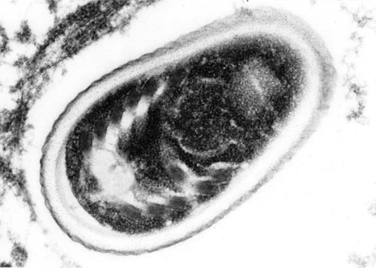
The infective form of microsporidia (E. cuniculi) is a resistant spore which can survive for a long time in the environment. The spore extrudes its polar tubule and infects the host cell. The spore injects the infective sporoplasm into the eukaryotic host cell through the polar tubule. Inside the cell, the sporoplasm undergoes extensive multiplication. This multiplication occurs either by merogony (binary fission) or schizogony (multiple fission). Microsporidia develop by sporogony to mature spores in the cytoplasm or inside parasitophorous vacule. During sporogony, a thick wall is formed around the spore. The thick wall formed provides resistance to adverse environmental conditions. Once the spores increase in number and completely fill the cytoplasm of the host's cell, the cell membrane is disrupted and releases the spores to the surroundings. These free mature spores can infect new cells thus continuing the cycle.
Transmission
The majority of rabbits seem to become infected with E. cuniculi from their mothers. Infection can also occur when something contaminated by urine containing E. cuniculi spores has been ingested. Approximately one month after the rabbit is infected it begins to shed E. cuniculi spores via its urine. Spores continue to be shed for approximately 3 months, but can be shed throughout the rabbit's life.
Human-to-human transmission is also possible via transplantation of solid organs from an infected donor.
Neurological impairment
E. cuniculi is a fungi that commonly parasitizes rabbits. The majority of the time, these organisms do not cause any obvious clinical disease. In fact, most often, E. cuniculi remains dormant within rabbits, never causing any problems upon initial infection. These parasites rarely migrate to nervous tissue or other organs. On occasions when E. cuniculi do reach nervous tissue, rabbits can experience neurological impairment, characterized by partial or complete paralysis, loss of coordination, seizures, and head tilting. Neurological impairment is thought to occur when E. cuniculi multiply within the rabbit's body inducing the development of granulomas, which are believed to travel through the body via white blood cells. The only way to expel these parasites is via urine. Something to note, however, is that it is still unknown if the damage is incurred by E. cuniculi directly, through natural growth and replication, or by the immune response itself. This uncertainty arises because there have been many well documented diseases in which humans and animals' own immune systems react to a non-threatening pathogen, but the immune response itself is responsible for the disease.
Detection
Antibodies to E. cuniculi can be detected through serology (a blood test). A rabbit who has been exposed to E. cuniculi will produce antibodies as a result of this exposure. Serology is used to detect the level of the antibody to the organism in the blood (this result is known as the titre). A positive titre tells you that the rabbit has been exposed to this parasite at some point in its life. Note that this does not allow one to differentiate between simple exposure and an active infection. By performing serology (with an interval of time in between) and comparing the results, it is possible to determine if the rabbit is mounting an immune response to an active infection or if it was previously infected. However, even with a rising titre, one can't determine for certain that a particular health problem is being caused by E.cuniculi or something else. Head tilt, for example, has many causes other than E. cuniculi, including bacterial or viral infections, trauma and toxicity. A false negative titre may also result if an infected animal produces an inadequate amount of antibodies.
History
Microsporidia are obligate intracellular parasitic fungi infecting many animal groups. Lacking mitochondria and peroxysomes, these unicellular eukaryotes were first considered a deeply branching protist lineage that diverged before the endosymbiotic event that led to mitochondria. The discovery of a gene for a mitochondrial-type chaperone combined with molecular phylogenetic data later implied that microsporidia are atypical fungi that lost mitochondria during evolution. Its genome consists of approximately 2.9-megabases (Mbs) in 11 chromosomes, with a total of 1,997 potential protein-coding genes. Genome compaction is reflected by reduced intergenic spacers and by the shortness of most putative proteins relative to their eukaryote orthologues. The strong host dependence is illustrated by the lack of genes for some biosynthetic pathways and for the tricarboxylic acid cycle. Phylogenetic analysis lends substantial credit to the fungal affiliation of microsporidia. Because the E. cuniculi genome contains genes related to some mitochondrial functions (for example, Fe-S cluster assembly), it is possible that microsporidia have retained a mitochondrion-derived organelle.