
 | ||
Elbow dysplasia is a condition involving multiple developmental abnormalities of the elbow-joint in the dog, specifically the growth of cartilage or the structures surrounding it. These abnormalities, known as 'primary lesions', give rise to osteoarthritic processes. Elbow dysplasia is a common condition of certain breeds of dogs.
Contents
- The disease
- Causes
- Diagnosis and treatment
- Non surgical treatment
- BioScaffold Implant Procedure
- Total elbow replacement
- Grading for breeding purposes
- References
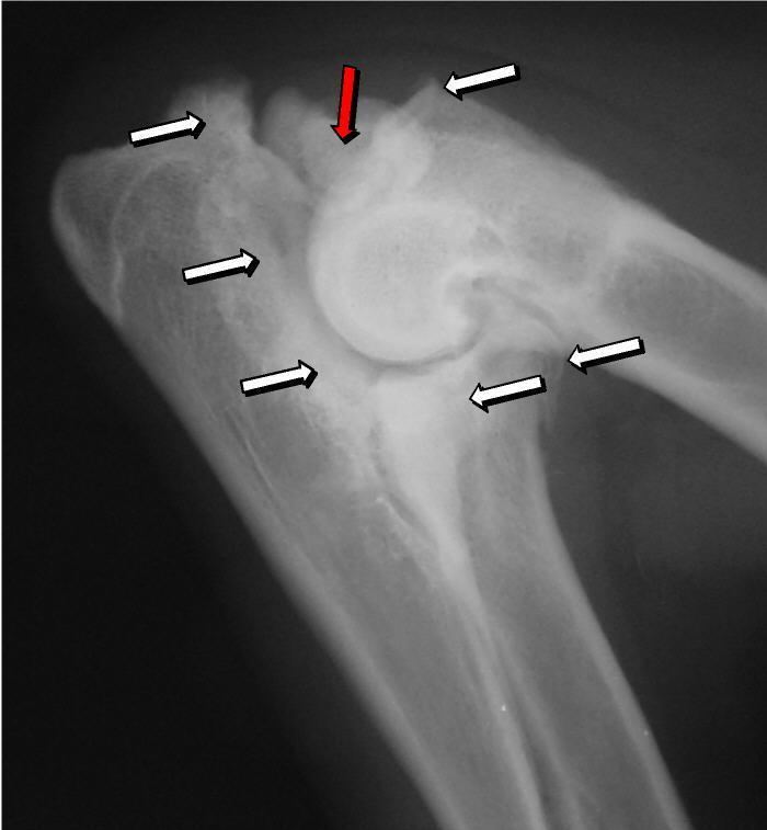
Most primary lesions are related to osteochondrosis, which is a disease of the joint cartilage and specifically Osteochondritis dissecans (OCD or OD), the separation of a flap of cartilage on the joint surface. Other common causes of elbow dysplasia included ununited anconeal process (UAP) and fragmented or ununited medial coronoid process (FCP or FMCP).
Osteochondritis dissecans is difficult to diagnose clinically as the animal may only exhibit an unusual gait. Consequently, OCD may be masked by, or misdiagnosed as, other skeletal and joint conditions such as hip dysplasia. The problem develops in puppyhood although often subclinically, and there may be pain or stiffness, discomfort on extension, or other compensating characteristics. Diagnosis generally depends on X-rays, arthroscopy, or MRI scans. While cases of OCD of the stifle go undetected and heal spontaneously, others are exhibited in acute lameness. Surgery is recommended once the animal has been deemed lame, before then non-surgical control is usually used.
The disease
Elbow Dysplasia is a significant genetically determined problem in many breeds of dog, often manifesting from puppyhood and continuing for life. In elbow dysplasia, the complex elbow joint suffers from a structural defect, often related to its cartilage. This initial condition, known as a "primary lesion", causes an abnormal level of wear and tear and gradual degradation of the joint, at times disabling or with chronic pain. Secondary processes such as inflammation and osteoarthritis can arise from this damage which increase the problem and add further problems of their own.
Causes
The most common cause is osteochondrosis, which is a disease of the joint cartilage, and specifically Osteochondritis dissecans (OCD or OD), the separation of a flap of cartilage from the joint surface as a result of avascular necrosis, which in turn arises from failed blood flow in the subchondral bone. Other common causes of elbow dysplasia included ununited anconeal process (UAP) and fractured or ununited medial coronoid process (FCP or FMCP).
In OCD, the normal change of cartilage to bone in the development of the joint fails or is delayed. The cartilage continues to grow and may split or become necrotic. The cause is uncertain, but possibly includes genetics, trauma, and nutrition (including excessive calcium and decreased Vitamin C intake). OCD lesions are found in the elbow at the medial epicondyle of the humerus. Specific conditions related to OCD include fragmentation of the medial coronoid process of the ulna (FMCP) and an ununited anconeal process of the ulna (UAP). All types of OCD of the elbow are most typically found in large breed dogs, with symptoms starting between the ages of 4 to 8 months. Males are affected twice as often as females. The disease often affects both elbows (30 to 70 percent of the time), and symptoms include intermittent lameness, joint swelling, and external rotation and abduction of the paw. Osteoarthritis will develop later in most cases.
UAP is caused by a separation from the ulna of the ossification center of the anconeal process. FMCP is caused by a failure of the coronoid process to unite with the ulna. OCD of the medial epicondyle of the humerus is caused by disturbed endochondral fusion of the epiphysis of the medial epicondyle with the distal end of the humerus, which may in turn be caused by avulsion of the epiphysis.
Diagnosis and treatment
Diagnosis is through x-rays, arthroscopy or CT (computed tomography). In cases with significant lameness, surgery is the best option, especially with UAP. However, conservative treatment is often enough for cases of FMCP and OCD of the medial humeral epicondyle. The dogs are exercised regularly and given pain medication, and between the ages of 12 to 18 months the lameness will often improve or disappear. Control of body weight is important in all cases of elbow dysplasia, and prevention of quick growth spurts in puppies may help to prevent the disease.
Surgery for FMCP consists of removal of cartilage and bone fragments and correction of any incongruity of the joint. Reattachment of UAP with a screw is usually attempted before the age of 24 weeks, and after that age the typical treatment is removal of the UAP. Without surgery, UAP rapidly progresses to osteoarthritis, but with FMCP osteoarthritis typically occurs with or without surgery. Osteoarthritis is also a common sequela of OCD of the humerus despite medical or surgical treatment. Elbow replacement surgery has been developed and can be an option for treatment
Non-surgical treatment
Conservative therapies include NSAIDs, pain medication, weight management and exercise restriction. The problems with these therapies is that they do not work well, especially long-term.
BioScaffold Implant Procedure
In a recent comparative orthopedic study, a new bioscaffold having an embryonic-like structure has shown positive clinical outcomes in dogs with advanced, end stage osteoarthritis. The bioscaffold was implanted into intra-articular areas and reported up to 90-days of clinical improvement after a single implant. The bioscaffold has been shown to cause infiltrating cells to upregulate a variety of tissue repair factors including aggrecan, connective tissue growth factor, bone morphogenetic protein, transforming growth factors, and other tissue repair factors associated with osteoarthritis [1] TR BioSurgical, LLC.
Total elbow replacement
The elbow is a complex joint, bears 60% of body load, and tolerates problems less well than the hips. As a result, elbow replacement is more complex than hip replacement, rehabilitation can take significantly longer, and some degree of lameness will remain. The surgery is classified as of 2010 as a "salvage" operation - that is, a last resort for an otherwise viable animal. Success rates of around 80 - 85% (approximately 5 in 6) were being discussed by sources in 2005 and again in 2008.
There are also fewer options if the replacement fails, the main option being arthrodesis (surgical fusion of the joint) which results in a pain-free but lame gait. However arthrodesis is itself a complex surgery with a long recovery time, and if arthrodesis is required, additional strain will be borne by other nearby joints, so other leg and shoulder conditions such as osteoarthritis may become more significant.
In 2012, Dr. Kirk Wendelburg, DVM, founder of Animal Specialty Group (ASG) in Los Angeles, collaborated with Swiss veterinary technology company, Kyon, to design an advanced elbow replacement system. This new system maintains the normal range of motion through the sagittal plane, around a normal center of rotation, while allowing normal supination and pronation. The compact design has no mechanical linkages between the elbow compartments, significantly reducing undue stresses which may lead to early failure. Dr. Wendelburg also invented the implantational procedure for the prosthesis. This repeatable, highly advanced procedure also allows the surgeon to make an intraoperative decision on whether a total or partial elbow replacement is needed. For example, a dog with only medial compartment disease may only receive a partial replacement. This intraoperative flexibility is possible due to the modular design of the implant. The system invented by Dr. Wendelburg is the first and only to allow for a, "Biomechanically Anatomical, Nonconstrained, Compartmental (BANC) Elbow Arthroplasty."
Grading for breeding purposes
The Orthopedic Foundation for Animals in the United States will grade elbow X-rays of dogs intended for breeding.