
ICD-9-CM 756.83 MedlinePlus 001468 | ICD-10 Q79.6 (ILDS Q82.817) DiseasesDB 4131 eMedicine derm/696 ped/654 | |
 | ||
Ehlers–Danlos syndrome (EDS) is a group of genetic connective tissue disorders. Symptoms can vary from mildly loose joints to life-threatening complications such as aortic dissection. Chronic pain or early osteoarthritis may also occur.
Contents
- Signs and symptoms
- Musculoskeletal
- Skin
- Cardiovascular
- Other manifestations or complications
- Genetics
- Diagnosis
- Classification
- Other types
- Differential diagnosis
- Management
- Surgery
- Prognosis
- Epidemiology
- Society and culture
- Other species
- References
EDS is caused by a defect in the structure, production, or processing of collagen or proteins that interact with collagen. The collagen in connective tissue helps tissues resist deformation. Collagen is an important contributor to the physical strength of tissue; abnormal collagen renders these structures more elastic. In some cases, it can be life-threatening. People with joint pain may be misdiagnosed with hypochondriasis, depression, chronic fatigue syndrome, or other conditions. There may be poor knowledge about EDS among practitioners.
There is no known cure for EDS. Treatment is supportive, including close monitoring of the digestive, excretory, and particularly the cardiovascular systems. Physical therapy, bracing, and corrective surgery may help with injuries and pain that tend to develop in certain types of EDS, although extra caution and special practices are advised to prevent permanent damage. EDS is a long term syndrome.
EDS affects about 1 in 5,000 people globally. Excess mobility was first described by Hippocrates in 400 BC. The syndrome is named after two physicians, Edvard Ehlers from Denmark and Henri-Alexandre Danlos from France, who described it at the turn of the 20th century.
Signs and symptoms
Signs vary widely based on which type of EDS the patient has. In each case, however, the signs are ultimately due to faulty or reduced amounts of collagen. EDS typically affects the joints, skin, and blood vessels. Following is a list of major signs and symptoms.
Musculoskeletal
Skin
Cardiovascular
Other manifestations or complications
Because it is often undiagnosed or misdiagnosed in childhood, some instances of Ehlers–Danlos syndrome have been mischaracterized as child abuse.
The pain associated with this condition is a serious complication.
Genetics
As mentioned under "Classification" above, only some variations of Ehlers-Danlos can be positively identified as tied to specific genetic variation.
Mutations in the following genes can cause subtypes of the Ehlers–Danlos syndrome:
Mutations in these genes usually alter the structure, production, or processing of collagen or proteins that interact with collagen. Collagen provides structure and strength to connective tissue. A defect in collagen can weaken connective tissue in the skin, bones, blood vessels, and organs, resulting in the features of the disorder.
Inheritance patterns depend on the type of Ehlers–Danlos syndrome. Most forms of the condition are inherited in an autosomal dominant pattern, which means only one of the two copies of the gene in question must be altered to cause the disorder. The minority are inherited in an autosomal recessive pattern, which means both copies of the gene must be altered for a person to be affected by the condition. It can also be an individual (de novo or "sporadic") mutation. Refer to the summary for each type of Ehlers–Danlos syndrome for a discussion of its inheritance pattern.
Diagnosis
A diagnosis can be made by an evaluation of medical history and clinical observation. The Beighton criteria is widely used to assess the degree of joint hypermobility. DNA and biochemical studies can help identify affected individuals. Diagnostic tests include collagen gene mutation testing, collagen typing via skin biopsy, echocardiogram, and lysyl hydroxylase or oxidase activity. However, these tests are not able to confirm all cases, especially in instances of an unmapped mutation, so clinical evaluation by a geneticist remains essential. If there are multiple affected individuals in a family, it may be possible to perform prenatal diagnosis using a DNA information technique known as a linkage study.
Classification
Up until 1997, the classification system for EDS included 10 specific types and also acknowledged that other extremely rare types existed. At this time, the classification system underwent an overhaul and was reduced to 6 major types using descriptive titles. Genetic specialists recognize that other types of this condition exist, but have only been documented in single families. Except for Hypermobility (type 3), some of the specific mutations involved have been identified and they can be precisely identified by genetic testing; this is valuable due to a great deal of variation in individual cases. However, negative genetic test results do not rule out the diagnosis, since not all of the mutations have been discovered; therefore the clinical presentation is very important. Although the classifications are well defined, it is rare for a case to fit neatly in a single category, and cross-over symptoms lead to under-diagnosis or misdiagnosis. Therefore, patients should not rely on the "fact" of having a certain type of EDS if cross-over symptoms are evident because of possibly life-threatening symptoms. For example, it is possible for an individual with Classical EDS to exhibit symptoms of Hypermobility or Vascular EDS.
In decreasing order of prevalence in the population, the classifications are:
Other types
Forms of EDS in this category may present with soft, mildly stretchable skin, shortened bones, chronic diarrhea, joint hypermobility and dislocation, bladder rupture, or poor wound healing. Inheritance patterns in this group include X-linked recessive, autosomal dominant, and autosomal recessive. Examples of types of related syndromes other than those above reported in the medical literature include:
Differential diagnosis
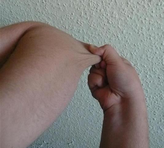
There are several disorders that share some characteristics with Ehlers–Danlos syndrome. For example, in cutis laxa the skin is loose, hanging, and wrinkled. In EDS, the skin can be pulled away from the body but is elastic and returns to normal when let go. In Marfan syndrome, the joints are very mobile and similar cardiovascular complications occur. People with EDS tend to have a "Marfanoid" appearance (e.g., tall, skinny, long arms and legs, "spidery" fingers). However, physical appearance and features in several types of Ehlers-Danlos Syndrome also have characteristics including short stature, large eyes, and the appearance of a small mouth and chin, due to a small palate. The palate can have a high arch, causing dental crowding. Blood vessels can sometimes be easily seen through translucent skin, especially on the chest. In the past, Menkes disease, a copper metabolism disorder, was thought to be a form of Ehlers–Danlos syndrome. It is not uncommon for patients to be misdiagnosed with fibromyalgia, bleeding disorders or other disorders that can mimic EDS symptoms before a correct diagnosis is made. Because of these similar disorders and complications that can arise from an unmonitored case of EDS, a correct diagnosis is very important. Pseudoxanthoma elasticum (PXE) is worth consideration in diagnosing a patient.
Management
There is no known cure for Ehlers Danlos Syndrome. Treatment is palliative. Close monitoring of the cardiovascular system, physiotherapy, occupational therapy, and orthopedic instruments (e.g., wheelchairs, bracing, casting) may be helpful. Orthopedic instruments are helpful for the prevention of further joint damage, especially for long distances, although it is advised that individuals not become entirely dependent on them until there are no other options for mobility. One should avoid activities that cause the joint to lock or overextend.
A physician may prescribe casting to stabilize joints. Physicians may refer a patient to an orthotist for orthotic treatment (bracing). Physicians may also consult a physical and/or occupational therapist to help strengthen muscles and to teach people how to properly use and preserve their joints.
There are different types of physiotherapy. Aquatic therapy promotes muscular development and coordination. With manual therapy, the joint will be gently mobilized within the range of motion and/or manipulations. If conservative therapy is not helpful, surgical repair of joints may be necessary. Medication to decrease pain or manage cardiac, digestive, or other related conditions may be prescribed. To decrease bruising and improve wound healing, some patients have responded to ascorbic acid (vitamin C). Special precautions are often taken by medical care workers because of the sheer amount of complications that tend to arise in EDS patients. In Vascular EDS, signs of chest or abdominal pain are to be considered trauma situations.
In general, medical intervention is limited to symptomatic therapy. Before pregnancy, patients with EDS should have genetic counseling and familiarize themselves with the risks to their own bodies that pregnancy poses. Children with EDS should be provided with information about the disorder so they can understand why contact sports and other physically stressful activities should be avoided. Children should be taught early on that demonstrating the unusual positions they can maintain due to loose joints should not be done as this may cause early degeneration of the joints. Patients may find it hard to cope with the drawbacks of the disease. In this case, emotional support and behavioral and psychological therapy can be useful. Support groups can be immensely helpful for patients dealing with major lifestyle changes and poor health. Family members, teachers, and friends should be informed about EDS so they can accept and assist the child.
Surgery
The instability of joints, leading to (sub)luxations and joint pain, often require surgical intervention in patients with Ehlers–Danlos syndrome. Instability of almost all joints can happen but appear most often in the lower and upper extremities, with the wrist, fingers, shoulder, knee, hip, and ankle being most common.
Common surgical procedures are joint debridement, tendon replacements, capsulorraphy, and arthroplasty. Studies have shown that after surgery, degree of stabilization, pain reduction, and patient satisfaction can improve, but surgery does not guarantee an optimal result: Patients and surgeons report being dissatisfied with the results. Consensus is that conservative treatment is more effective than surgery, particularly since patients have extra risks of surgical complications due to the disease. Three basic surgical problems arise due to EDS: the strength of the tissues is decreased, which makes the tissue less suitable for surgery; the fragility of the blood vessels can cause problems during surgery; and wound healing is often delayed or incomplete. If considering surgical intervention, it would be prudent to seek care from a surgeon with extensive knowledge and experience in treating patients with EDS and joint hypermobility issues.
Studies have shown that local anesthetics, arterial catheters and central venous catheters cause a higher risk in haematoma formation in patients with Ehlers–Danlos syndrome. Ehlers-Danlos patients also show a resistance to local anaesthetics. Resistance to Xylocaine and Bupivacaine is not uncommon, and Carbocaine tends to work better in EDS patents. Special recommendations for anesthesia in EDS patients are prepared by orphan anesthesia.eu and deal with all aspects of anesthesia for EDS patients. Detailed recommendations for anesthesia and perioperative care of patients with EDS should be used to improve patient safety.
Surgery with Ehlers-Danlos patients requires careful tissue handling and a longer immobilization afterward.
Prognosis
The outlook for individuals with EDS depends on the type of EDS they have. Symptoms vary in severity, even within one sub-type, and the frequency of complications changes individually. Some people have negligible symptoms while others are severely restricted in their daily life. Extreme joint instability, chronic musculoskeletal pain, degenerative joint disease, frequent injuries, and spinal deformities may limit mobility. Severe spinal deformities may affect breathing. In the case of extreme joint instability, dislocations may result from simple tasks such as rolling over in bed or turning a doorknob. Secondary conditions such as autonomic dysfunction or cardiovascular problems, occurring in any type, can affect prognosis and quality of life. Severe mobility-related disability is seen more often in Hypermobility-type than in Classical-type or Vascular-type.
Although all types are potentially life-threatening, the majority of individuals will have a normal lifespan. However, those with blood vessel fragility have a high risk of fatal complications. Arterial rupture is the most common cause of sudden death in EDS. Spontaneous arterial rupture most often occurs in the second or third decade, but can occur at any time. The median life-expectancy in the population with Vascular EDS is 48 years.
EDS is a lifelong condition. Affected individuals may face social obstacles related to their disease daily. Some people with EDS have reported living with fear of significant and painful ruptures, their condition worsening, becoming unemployed due to physical and emotional burdens, and social stigmatization in general.
Epidemiology
Ehlers–Danlos syndrome is an inherited disorder estimated to occur in about 1 in 5,000 births worldwide. Initially, prevalence estimates ranged from 1 in 250,000 to 1 in 500,000 people, but these estimates were soon found to be vastly inaccurate as the disorder received further study and medical professionals became more adept at accurately diagnosing EDS. In fact, many experts now believe that Ehlers–Danlos syndrome may be far more common than the currently accepted estimate due to the wide range of severities with which the disorder presents. The prevalence of the six types differs dramatically. The most commonly occurring is the Hypermobility type, followed by the Classical type. The other types of Ehlers–Danlos syndrome are very rare. For example, fewer than 10 infants and children with the dermatosparaxis type have been described worldwide. Ehlers–Danlos affects males and females of all racial and ethnic backgrounds, although some types are more common among certain groups than others.
Society and culture
Other species
Ehlers–Danlos-like syndromes have been shown to be hereditary in Himalayan cats, some domestic shorthair cats, and in certain breeds of cattle. It is seen as a sporadic condition in domestic dogs.
Degenerative suspensory ligament desmitis (DSLD) is a similar condition seen in many breeds of horses. It was originally notated in the Peruvian Paso and thought to be a condition of overwork and older age. However, the disease is being recognized in all age groups and all activity levels. It has even been noted in newborn foals. The latest research has led to the renaming of the disease as equine systemic proteoglycan accumulation, after the possible systemic and hereditary components being delineated by the University of Georgia.