
MeSH A09.246.272.702 TA A15.3.01.052 | FMA 9595 | |
 | ||
Latin membrana tympanica; myringa | ||
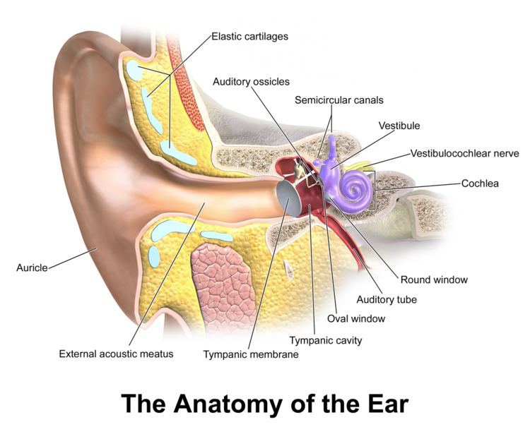
In the anatomy of humans and various other tetrapods, the eardrum, also called the tympanic membrane or myringa, is a thin, cone-shaped membrane that separates the external ear from the middle ear. Its function is to transmit sound from the air to the ossicles inside the middle ear, and then to the oval window in the fluid-filled cochlea. Hence, it ultimately converts and amplifies vibration in air to vibration in fluid. The malleus bone bridges the gap between the eardrum and the other ossicles.
Contents
- Structure
- Nerve supply
- Relations
- Umbo
- Rupture
- Surgical puncture for treatment of middle ear infections
- Society and culture
- References
Rupture or perforation of the eardrum can lead to conductive hearing loss. Collapse or retraction of the eardrum can cause conductive hearing loss or cholesteatoma.
Structure
There are two general regions of the eardrum: the pars flaccida in the upper region and the pars tensa. The pars flaccida consists of two layers, is relatively fragile, and is associated with eustachian tube dysfunction and cholesteatomas. The larger pars tensa region consists of three layers: skin, fibrous tissue, and mucosa. It is comparatively robust and is the region most commonly associated with perforations.
The pars tensa forms most of the tympanic membrane. Its periphery is thick and forms a fibrocartilaginous ring called the anulus tympanicus. The central part of the pars tensa is tented inward at the level of the tip of malleus and is called the umbo. When the eardrum is illuminated during an examination, a cone of light radiates from the tip of the malleus to the periphery in the antero-inferior quadrant. The pars flaccida is above the lateral process of the malleus between the notch of Rivinus and the anterior and posterior malleal folds. It appears slightly pinkish.
Nerve supply
Sensory innervation of the external surface of the tympanic membrane is mainly by the auriculotemporal nerve, a branch of the mandibular nerve [V3]. It also has contributions from the auricular branch of the vagus nerve [X], the facial nerve [VII] and a possible contribution from the glossopharyngeal nerve [IX]. Sensory innervation of the inner surface of the tympanic membrane is by the glossopharyngeal nerve [IX].
Relations
The tympanic membrane is superiorly related to middle cranial fossa, posteriorly to the ear ossicles and the facial nerve, inferiorly to the parotid gland and anteriorly to the temporomandibular joint.
Umbo
The umbo is the most depressed part of the tympanic membrane. The manubrium of the malleus is firmly attached to the medial surface of the membrane as far as its center, which it draws toward the tympanic cavity; the lateral surface of the membrane is thus concave, and the most depressed part of this concavity is named the umbo.
Rupture
An unintentional perforated eardrum (rupture) has been described in blast injuries during conflict and air travel, usually when the congestion of an upper respiratory infection has prevented equalization of pressure in the middle ear. It happens in sport and recreation, such as swimming, diving with a poor entry into the water, scuba diving, and martial arts. In the published literature, 80% to 95% have recovered completely without intervention in two to four weeks.
These injuries, even in a recreational or athletic setting, are blast injuries. Many will experience some short-lived hearing loss and ringing in the ear (tinnitus) but can be reassured that it, in all likelihood, will pass. A very few will experience temporary disequilibrium (vertigo). There may be some bleeding from the ear canal if the eardrum has been ruptured. Naturally, the foregoing reassurances become more guarded as the force of injury increases, as in military or combat situations.
Surgical puncture for treatment of middle ear infections
A myringotomy (tympanotomy, tympanostomy) is a surgical procedure in which a tiny incision is created in the eardrum to relieve pressure caused by excessive buildup of fluid, or to drain pus from the middle ear. The fluid or pus comes from a middle ear infection (otitis media), which is a common problem in children. A tympanostomy tube is inserted into the eardrum to keep the middle ear aerated for a prolonged time and to prevent reaccumulation of fluid. Without the insertion of a tube, the incision usually heals spontaneously in two to three weeks. Depending on the type, the tube is either naturally extruded in 6 to 12 months or removed during a minor procedure.
Those requiring myringotomy usually have an obstructed or dysfunctional eustachian tube that is unable to perform drainage or ventilation in its usual fashion. Before the invention of antibiotics, myringotomy without tube placement was also used as a major treatment of severe acute otitis media.
Society and culture
The Bajau people of the Pacific intentionally rupture their eardrums at an early age to facilitate diving and hunting at sea. Many older Bajau therefore have difficulties hearing.