
TA A10.1.02.428 | FMA 24230 | |
 | ||
Latin trigonum cystohepaticum | ||
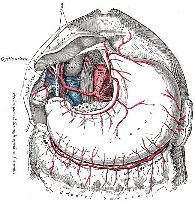
The hepatobiliary triangle (or cystohepatic triangle) is an anatomic space bordered by the cystic duct inferiorly, common hepatic duct medially and the inferior (visceral) surface of the liver superiorly. The cystic artery lies within the cystohepatic triangle, which is used to locate it during a laparoscopic cholecystectomy.
Contents
Eponym
Another name used to refer to this region is Calot's Triangle. It is named for Jean-François Calot. Of note, Calot's original description of the triangle in 1891 included the cystic duct, the common hepatic duct, and the cystic artery (not the inferior border of the liver as is commonly believed). The Hepatocystic triangle is the area bound by the cystic duct, common hepatic duct, and the liver margin.
Clinical significance
General surgeons frequently quiz medical students on this term and the name for the lymph node located within the triangle, Mascagni's lymph node or Lund's node, however many often erroneously refer to it as "Calot's node." The latter is frequently enlarged due to inflammation of the gallbladder (e.g. cholecystitis) or the biliary tract (e.g. cholangitis) and may be removed along with the gallbladder during surgical treatment (cholecystectomy).
Calot's triangle, containing the cystic artery, may also contain an accessory right hepatic artery or anomalous sectoral bile ducts. As a result dissection in the triangle of Calot is ill-advised until the lateral-most structures have been cleared and identification of the cystic duct is definitive. According to SESAP 12 (produced and distributed by the American College of Surgeons) dissection in the triangle of Calot is the most common cause of common bile duct injuries.