
 | ||
Circulating tumor cells (CTCs) are cells that have shed into the vasculature or lymphatics from a primary tumor and are carried around the body in the circulation. CTCs thus constitute seeds for the subsequent growth of additional tumors (metastases) in vital distant organs, triggering a mechanism that is responsible for the vast majority of cancer-related deaths.
Contents
- Types of CTCs
- Frequency of CTCs
- Clinical utility
- CellSearch method
- Epic sciences method
- Maintrac
- ISET Test
- Other methods
- Oncopheresis Machine dialysis
- Characterization of CTC
- Further characterisation of CTC
- Morphological definition
- References
CTCs were observed for the first time in 1869 in the blood of a man with metastatic cancer by Thomas Ashworth, who postulated that “cells identical with those of the cancer itself being seen in the blood may tend to throw some light upon the mode of origin of multiple tumours existing in the same person”. A thorough comparison of the morphology of the circulating cells to tumor cells from different lesions led Ashworth to conclude that “One thing is certain, that if they [CTC] came from an existing cancer structure, they must have passed through the greater part of the circulatory system to have arrived at the internal saphena vein of the sound leg”.
The importance of CTCs in modern cancer research began in the mid 1990s with the demonstration [J. Uhr, UT-Dallas, L. Terstappen and P. Liberti, Immunicon, Philadelphia] that CTC's exist early on in the course of the disease. Those results were made possible by exquisitely sensitive magnetic separation technology employing Ferrofluids (colloidal magnetic nanoparticles) and high gradient magnetic separators invented by Liberti at Immunicon and motivated by theoretical calculations by Liberti and Terstappen that indicated very small tumors shedding cells at less than 1.0% per day should result in detectable cells in blood. A variety of other technologies have been applied to CTC enumeration and identification since that time.
Modern cancer research has demonstrated that CTCs derive from clones in the primary tumor, validating Ashworth's remarks. The significant efforts put into understanding the CTCs biological properties have demonstrated the critical role circulating tumor cells play in the metastatic spread of carcinoma. Furthermore, highly sensitive, single-cell analysis demonstrated a high level of heterogeneity seen at the single cell level for both protein expression and protein localization and the CTCs reflected both the primary biopsy and the changes seen in the metastatic sites.
Tissue biopsies are poor diagnostic procedures: they are invasive, cannot be used repeatedly, and are ineffective in understanding metastatic risk, disease progression, and treatment effectiveness. CTCs thus could be considered a “liquid biopsy” which reveals metastasis in action, providing live information about the patient’s disease status. Analysis of blood samples found a propensity for increased CTC detection as the disease progressed in individual patients. Blood tests are easy and safe to perform and multiple samples can be taken over time. By contrast, analysis of solid tumors necessitates invasive procedures that might limit patient compliance. The ability to monitor the disease progression over time could facilitate appropriate modification to a patient's therapy, potentially improving their prognosis and quality of life. The important aspect of the ability to prognose the future progression of the disease is elimination (at least temporarily) of the need for a surgery when the repeated CTC counts are low and not increasing; the obvious benefits of avoiding the surgery include avoiding the risk related to the innate tumor-genicity of cancer surgeries. To this end, technologies with the requisite sensitivity and reproducibility to detect CTCs in patients with metastatic disease have recently been developed.
Types of CTCs
1. Traditional CTCs are confirmed cancer cells with an intact, viable nucleus; express cytokeratins, which demonstrate epithelial origin; have an absence of CD45, indicating the cell is not of hematopoietic origin; and are often larger cells with irregular shape or subcellular morphology.
2. Cytokeratin negative (CK-) CTCs are cancer stem cells or cells undergoing epithelial-mesenchymal transition (EMT). CK-CTCs may be the most resistant and most prone to metastasis; express neither cytokeratins nor CD45; have morphology similar to a cancer cell; and importantly have gene or protein expression or genomics associated with cancer.
3. Apoptotic CTCs are traditional CTCs that are undergoing apoptosis (cell death): Epic Sciences technology identifies nuclear fragmentation or cytoplasmic blebbing associated with apoptosis. Measuring the ratio of traditional CTC to apoptotic CTCs – from baseline to therapy – provides clues to a therapy’s efficacy in targeting and killing cancer cells.
4. Small CTCs are cytokeratin positive and CD45 negative, but with sizes and shapes similar to white blood cells. Importantly, small CTCs have cancer-specific biomarkers that identify them as CTCs. Small CTCs have been implicated in progressive disease and differentiation into small cell carcinomas, which often require a different therapeutic course.
5. CTC clusters are two or more individual CTCs bound together. The CTC cluster may contain traditional, small or CK- CTCs. These clusters have cancer-specific biomarkers that identify them as CTCs. Some studies have reported that these clusters are associated with increased metastatic risk and poor prognosis. However, one study has reported that contrary to existing consensus, at least a discrete population of these clusters are non-malignant, and derive instead from the tumor-endothelia. These circulating tumor-endothelial clusters also show epithelial-mesenchymal markers but do not mirror the genetics of the primary tumor.
Frequency of CTCs
The detection of CTCs may have important prognostic and therapeutic implications but because their numbers can be very small, these cells are not easily detected. It is estimated that among the cells that have detached from the primary tumor, only 0.01% can form metastases.
Circulating tumor cells are found in frequencies on the order of 1-10 CTC per mL of whole blood in patients with metastatic disease. For comparison, a mL of blood contains a few million white blood cells and a billion red blood cells, see figure 1. This low frequency, associated to difficulty of identifying cancerous cells, means that a key component of understanding CTCs biological properties require technologies and approaches capable of isolating 1 CTC per mL of blood, either by enrichment, or better yet with enrichment-free assays that identify all CTC subtypes in sufficiently high definition to satisfy diagnostic pathology image-quantity requirements in patients with a variety of cancer types. To date CTCs have been detected in several epithelial cancers (breast, prostate, lung, and colon) and clinical evidences indicate that patients with metastatic lesions are more likely to have CTCs isolated.
CTCs are usually (in 2011) captured from the vasculature by using specific antibodies able to recognize specific tumoral marker (usually EpCAM); however this approach is biased by the need for a sufficient expression of the selected protein on the cell surface, event necessary for the enrichment step. Moreover, since EpCAM and other proteins (e.g. cytokeratins) are not expressed in some tumors and can be down regulated during the epithelial to mesenchymal transition (EMT), new enrichment strategies are required.
First evidence indicates that CTC markers applied in human medicine are conserved in other species. Five of the more common markers including CK19 are also useful to detect CTC in the blood of dogs with malignant mammary tumors.
Newer approaches are able to identify more cells out 7.5 ml of blood, like isoflux or maintrac.
Clinical utility
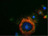
To date, a variety of research methods have been developed to isolate and enumerate CTC. The only U.S. Food and Drug Administration (FDA) cleared methodology for enumeration of CTC in whole blood is the CellSearch system. Extensive clinical testing done using this method shows that presence of CTC is a strong prognostic factor for overall survival in patients with metastatic breast, colorectal or prostate cancer, see figure 2
CellSearch method
This method is based on the use of iron nano-particles coated with a polymer layer carrying biotin analogues and conjugated with antibodies anti EpCAM for capturing CTCs, and on the use of an analyzer to take images of isolated cells upon their staining with specific fluorescent antibody conjugates. Blood is sampled in an EDTA tube with an added preservative. Upon arrival in the lab, 7.5mL of blood is centrifuged and placed in a preparation system. This system first enriches the tumor cells immunomagnetically by means of ferrofluid nano-particles and a magnet. Subsequently recovered cells are permeabilized and stained with a nuclear stain, a fluorescent antibody conjugate against CD45 (leukocyte marker), and cytokeratin 8, 18 and 19 (CKs). The sample is then scanned on an analyzer which takes images of the nuclear, cytokeratin, and CD45 stains. To be considered a CTC a cell must contain a nucleus, be positive for cytoplasmic expression of cytokeratin as well as negative for the expression of CD45 marker, and have a diameter larger than 5 µm. If the total number of tumor cells found to meet the criteria cited above is 5 or more, a blood sample is positive. In studies done on prostate, breast and colon cancer patients, median survival of metastatic patients with positive samples is about half the median survival of metastatic patients with negative samples. This system is characterized by a recovery capacity of 93% and a detection limit of one CTC per 7.5 mL of whole blood. Despite its sensitivity and reproducibility, the CellSearch method requires specific equipment to perform the analysis.
Epic sciences method
This method involves technology to separate nucleated cells from red blood cells, which lack a nucleus. All nucleated cells, including normal white blood cells and CTCs, are exposed to fluorescent-tagged antibodies specific for cancer biomarkers. In addition, Epic’s imaging system captures pictures of all the cells on the slide (approximately 3 million), records the precise coordinates of each cell, and analyzes each cell for 90 different parameters, including the fluorescence intensity of the four fluorescent markers and 86 different morphological parameters. Epic can also use FISH and other staining techniques to look for abnormalities such as duplications, deletions, and rearrangements. The imaging and analysis technology also allows for the coordinates of every cell on a slide to be known so that a single cell can be retrieved from the slide for analysis using next-generation sequencing. A hematopathology-trained algorithm incorporates numerous morphology measurements as well as expression from cytokeratin and CD45. The algorithm then proposes candidate CTCs that a trained reader confirms. Cells of interest are analyzed for relevant phenotypic and genotypic markers, with regional white blood cells included as negative controls. Epic’s molecular assays measure protein expression and also interrogate genomic abnormalities in CTCs for more than 20 different cancer types.
Maintrac
Maintrac is a diagnostic platform applying microscopic methods to identify rare cells in body fluids and their molecular characteristics.
Concerning circulating tumor cells, maintrac is using an approach based on microscopic identification of circulating tumor cells. To prevent damage and loss of the cells during the process, maintrac uses just two steps towards the identification. In contrast to many other methods, maintrac does not purify the cells or enriches them, but identifies them within the context of the other blood compounds. To obtain vital cells and to reduce stress of those cells, blood cells are prepared by only one centrifugation step and erythrocyte lysis. Like CellSearch maintrac uses an EpCAM antibody. It is, however, not used for enrichment but rather as a fluorescent marker to identify those cells. Together with the nuclear staining with propidium iodide the maintrac method can distinguish between dead and living cells. Only vital, propidium excluding EpCAM positive cells are counted as potential tumor cells. Only living cells can grow into tumors, therefore dying EpCAM positive cells can do no harm. The suspension is analysed by fluorescence microscopy, which automatically counts the events. Simultaneous event galleries are recorded to verify whether the software found a true living cell and to differentiate between skin epithelial cells for example. Close validation of the method showed that additional antibodies of cytokeratins or CD45 did not have any advantage.
Unlike other methods maintrac does not use the single cell count as a prognostic marker, rather maintrac utilizes the dynamics of the cell count. Rising tumor cell numbers are an important factor that tumor activity is ongoing. Decreasing cell counts are a sign for a successful therapy.
Therefore, maintrac can be used in following situations:
Circulating tumor cells as early diagnosis for cancer recurrence:
Studies showed that under certain circumstances also EpCAM positive cells can be found in the blood. Inflammation diseases like Morbus Crohn or Colitits Ulcerosa also show increased levels of EpCAM positive cells. Patients with severe skin burns can also carry EpCAM positive cells in the blood, which may falsify results. So use of EpCAM positive cells as a tool for early diagnosis is not recommended.
ISET Test
The ISET Test (Isolation by Size of Tumor cells / Trophoblastic cells) is a diagnostic blood test that detects circulating tumor cells in a blood sample. The test uses an in-vitro diagnostic system developed at INSERM, the Université Paris Descartes and Assistance Publique Hôpitaux de Paris (AP-HP) in order to isolate cancer cells from blood without loss and identify them through a diagnostic cytopathology-based approach.
Other methods
CTCs are pivotal to understanding the biology of metastasis and promise potential as a biomarker to noninvasively evaluate tumor progression and response to treatment. However, isolation and characterization of CTCs represent a major technological challenge, since CTCs make up a minute number of the total cells in circulating blood, 1–10 CTCs per mL of whole blood compared to a few million white blood cells and a billion red blood cells. Therefore, the major challenge for CTC researchers is the prevailing difficulty of CTC purification that allows the molecular characterization of CTCs. Several methods have been developed to isolate CTCs in the peripheral blood and essentially fall into two categories: biological methods and physical methods.
Another approach is the in vivo capture of CTCs of GILUPI GmbH. An antibody coated metal wire is inserted into a peripheral vein and stays there for a defined period (30 min). During this time, CTCs from the blood can bind to the antibodies (currently anti-EpCAM). After the incubation time, the wire is removed, washed and the native CTCs, isolated from the blood of the patient, can be further analysed. Molecular genetics as well as immunofluorescent staining and several other methods are possible. Advantage of this method is the higher blood volume that can be analysed for CTCs (approx. 750 ml in 30 min compared to 7.5 ml of a drawn blood sample). A sensitive double gradient centrifugation and magnetic cell sorting detection and enumeration method has been used to detect circulating epithelial cancer cells in breast cancer patients by a novel negative selection method. A similar methodology was used to detect circulating prostate cancer cells.
Oncopheresis (Machine dialysis)
The normal cellular components of blood principally consist of red blood cells (RBCs), leukocytes (white blood cells, or WBCs) and platelets. CTCs are abnormal blood cell components shed from tumors, and are invariably collected from a patient’s peripheral blood even though they are present throughout the circulatory system. Since all blood cells must pass through extremely narrow capillaries as they circulate through the body, CTCs in peripheral blood are found in extremely low numbers because they must circulate and survive through an obstacle course of harsh conditions, such as the heart, lungs and capillaries before they can appear in the peripheral blood. Aside from size and number, another important characteristic of blood cells is their deformability: the ability to change shape in order to traverse through the narrow capillaries which separate arterial blood (blood pumped out by the heart) from venous blood (blood returning to the heart after passing through a capillary). RBCs are known to be highly deformable, effectively and very quickly changing shape from a donut to a sausage as they pass through capillaries. Platelets and WBCs are less deformable than RBCs; but they nevertheless must and do traverse through capillaries, albeit at a slower rate. CTCs are the least deformable type of blood cells by several orders of magnitude. So far there are no approved methods, but Viatar CTC Solutions is undergoing human clinical tests through a specific mechanical filtration system in a 4-hr treatment for full body dialysis of CTCs from the blood.
Characterization of CTC
Any useful method for isolation of CTCs must allow (i) their identification and enumeration and (ii) their characterization through immunocytochemistry, fluorescence in situ hybridization (FISH) DNA and RNA assays, and all other relevant molecular techniques using DNA and RNA. When circulating tumor cells are captured from blood using filtration devices (such as ScreenCell isolation device), further morphological and molecular characterization is required to reveal important predictive information and report changes in CTC biology, for example during tumor relapse. ViewRNA assay for CTCs characterization is the only in situ hybridization technology that allows multiplex, single-molecule RNA detection of any RNA target. The exceptional sensitivity and specificity is achieved by using proprietary probe design, simultaneous branched DNA (bDNA) signal amplification and background suppression.
The captured CTCs on the filter membrane of a ScreenCell isolation device, are transferred to a 24-well cell culture plate for enumeration/characterization using ViewRNA ISH Cell Assay. A target-specific probe set containing 20 oligonucleotide pairs hybridizes to the target RNA. An oligo pair hybridization event is essential for support of the signal amplification structure, which is assembled by a series of sequential hybridization steps. Each fully assembled amplification structure is contained within 40−50 bp of target RNA with the capacity for 400-fold signal amplification. Therefore, a typical target-specific probe set (containing 20 oligo pairs) can generate 8,000-fold signal amplification at the location of the target RNA.
Further characterisation of CTC
Some drugs are particularly effective against cancers which fit certain requirements. For example, Herceptin is very effective in patients who are Her2 positive, but much less effective in patients who are Her2 negative. Once the primary tumor is removed, biopsy of the current state of the cancer through traditional tissue typing is not possible anymore. Often tissue sections of the primary tumor, removed years prior, are used to do the typing. Further characterisation of CTC may help determining the current tumor phenotype. FISH assays has been performed on CTC to as well as determination of IGF-1R, Her2, Bcl-2, [ERG (gene)|ERG], PTEN, AR status using immunofluorescence. Single cell level qPCR can also be performed with the CTCs isolated from blood.
Morphological definition
Morphological appearance is judged by human operators and is therefore subject to large inter operator variation. Several CTC enumeration methods exist which use morphological appearance to identify CTC, which may also apply different morphological criteria. A recent study in prostate cancer showed that many different morphological definitions of circulating tumor cells have similar prognostic value, even though the absolute number of cells found in patients and normal donors varied by more than a decade between different morphological definitions.