
 | ||
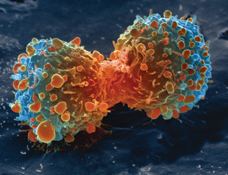
Cancer cells are cells that divide relentlessly, forming solid tumours or flooding the blood with abnormal cells. Cell division is a normal process used by the body for growth and repair. A parent cell divides to form two daughter cells, and these daughter cells are used to build new tissue, or to replace cells that have died as a result of ageing or damage. Healthy cells stop dividing when there is no longer a need for more daughter cells, but cancer cells continue to produce copies. They are also able to spread from one part of the body to another in a process known as metastasis.
Contents
Classification
There are different categories of cancer cell, defined according to the cell type from which they originate.
Histology
Cancer cells have distinguishing histological features visible under the microscope. The nucleus is often large and irregular, and the cytoplasm may also display abnormalities.
Nucleus
The shape, size, protein composition, and texture of the nucleus are often altered in malignant cells. The nucleus may acquire grooves, folds or indentations, chromatin may aggregate or disperse, and the nucleolus can become enlarged. In normal cells, the nucleus is often round or ellipsoid in shape, but in cancer cells the outline is often irregular. Different combinations of abnormalities are characteristic of different cancer types, to the extent that nuclear appearance can be used as a marker in cancer diagnostics and staging.
Causes
Cancer cells are created when the genes responsible for regulating cell division are damaged. Carcinogenesis is caused by mutation and epimutation of the genetic material of normal cells, which upsets the normal balance between proliferation and cell death. This results in uncontrolled cell division and the evolution of those cells by natural selection in the body. The uncontrolled and often rapid proliferation of cells can lead to benign or malignant tumours (cancer). Benign tumors do not spread to other parts of the body or invade other tissues. Malignant tumors can invade other organs, spread to distant locations (metastasis) and become life-threatening.
More than one mutation is necessary for carcinogenesis. In fact, a series of several mutations to certain classes of genes is usually required before a normal cell will transform into a cancer cell.
Damage to DNA can be caused by exposure to radiation, chemicals, and other environmental sources, but mutations also accumulate naturally over time through uncorrected errors in DNA transcription, making age another risk factor. Oncoviruses can cause certain types of cancer, and genetics are also known to play a role.
Stem cell research suggests that too much SP2 protein MAY turn stem cells into cancer cells. However, a lack of particular co-stimulated molecules that aid in the way antigens react with lymphocytes can impair the natural killer cells' function, ultimately leading to cancer.
Pathology
Immune system cells, such as White Blood cells are thought to use a dual receptor system when they determine whether or not to kill sick, or damaged human cells. If a cell is under stress, turning into tumors, or infected, molecules including MIC-A and MIC-B are produced to put on the surface of the cell. These work to help the white blood cells to detect and kill cancer cells.
Discovery
Cancer is an ancient disease with descriptions dating back to ancient Egypt. In 2016, a 1.7 million year old osteosarcoma was reported, representing the oldest documented malignant hominin cancer.
The understanding of cancer was significantly advanced during the Renaissance period and in to the Age of Discovery. Sir Rudolf Virchow, a German biologist and politician, studied microscopic pathology, and linked his observations to illness. He is described as "the founder of cellular pathology". In 1845, Virchow and John Hughes Bennett independently observed abnormal increase in white blood cells in patients. Virchow correctly identified the condition as blood disease, and named it leukämie in 1847 (later anglicised to leukemia). In 1857, he was the first to describe a type of tumour called chordoma that originated from the clivus (at the base of the skull).
Telomerase
Cancer cells have unique features that make them "immortal" according to some researchers. The enzyme telomerase is used to extend the cancer cell's life span. While the telomeres of most cells shortens after each division eventually causing the cell to die, telomerase extends the cell's telomeres. This is a major reason that cancer cells can accumulate over time creating tumors.
Cancer stem cells and drug resistance
Scientists have discovered a molecule on the surface of tumors that appears to promote drug resistance—by converting the tumor cells back into a stem cell-like state. When the tumor cells began to exhibit drug resistance, the cells were simultaneously transforming into a stem cell-like state, which made them impervious to the drugs. It appeared that the treatment itself was driving this transformation by activating a specific molecular pathway. Luckily, several existing drugs, such as Bortezomib for example, can attack this pathway and reverse the cellular transformation, thus ‘re-sensitizing’ the tumor to treatment.