
MeSH A07.231.114.228.351 FMA 50454 | TA A12.2.07.080 | |
 | ||
Latin Circulus arteriosus cerebriCirculus Willisi | ||
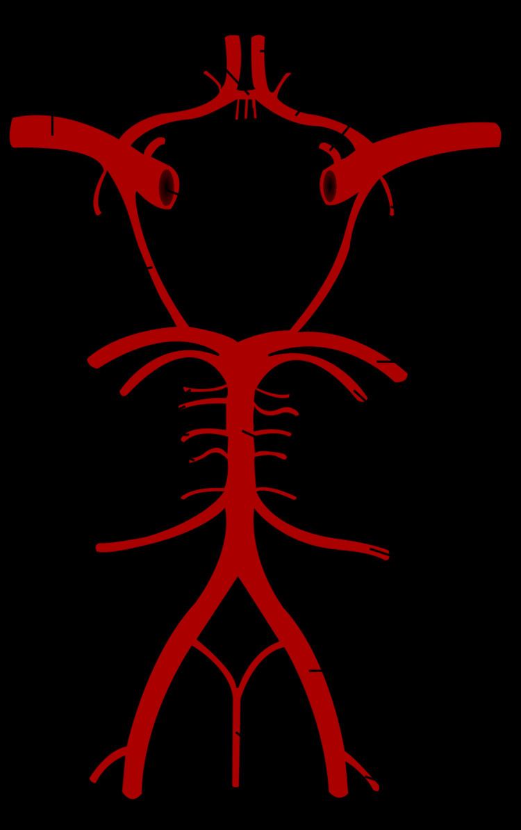
The circle of Willis (also called Willis' circle, loop of Willis, cerebral arterial circle, and Willis polygon) is a circulatory anastomosis that supplies blood to the brain and surrounding structures. It is named after Thomas Willis (1621–1675), an English physician.
Contents
Structure
The circle of Willis is a part of the cerebral circulation and is composed of the following arteries:
The middle cerebral arteries, supplying the brain, are not considered part of the circle.
Origin of arteries
The left and right internal carotid arteries arise from the left and right common carotid arteries.
The posterior communicating artery is given off as a branch of the internal carotid artery just before it divides into its terminal branches - the anterior and middle cerebral arteries. The anterior cerebral artery forms the anterolateral portion of the circle of Willis, while the middle cerebral artery does not contribute to the circle.
The right and left posterior cerebral arteries arise from the basilar artery, which is formed by the left and right vertebral arteries. The vertebral arteries arise from the subclavian arteries.
The anterior communicating artery connects the two anterior cerebral arteries and could be said to arise from either the left or right side.
All arteries involved give off cortical and central branches. The central branches supply the interior of the circle of Willis, more specifically, the Interpeduncular fossa. The cortical branches are named for the area they supply. Since they do not directly affect the circle of Willis, they are not dealt with here.
Variation
Considerable anatomic variation exists in the circle of Willis. Based on a study of 1413 brains, the classic anatomy of the circle is only seen in 34.5% of cases. In one common variation the proximal part of the posterior cerebral artery is narrow and its ipsilateral posterior communicating artery is large, so the internal carotid artery supplies the posterior cerebrum. In another variation the anterior communicating artery is a large vessel, such that a single internal carotid supplies both anterior cerebral arteries.
Function
The arrangement of the brain's arteries into the circle of Willis creates redundancy (analogous to engineered redundancy) for collateral circulation in the cerebral circulation. If one part of the circle becomes blocked or narrowed (stenosed) or one of the arteries supplying the circle is blocked or narrowed, blood flow from the other blood vessels can often preserve the cerebral perfusion well enough to avoid the symptoms of ischemia.
Subclavian steal syndrome
The redundancies that the circle of Willis introduce can also lead to reduced cerebral perfusion. In subclavian steal syndrome, blood is "stolen" from the circle of Willis to preserve blood flow to the upper limb. Subclavian steal syndrome results from a proximal stenosis (narrowing) of the subclavian artery, an artery supplied by the aorta which is also the same blood vessel that eventually feeds the circle of Willis via the vertebral artery.