
Days 24 | MeSH Chorionic+Villi | |
 | ||
Chorionic villi are villi that sprout from the chorion to provide maximum contact area with maternal blood.
Contents
They are an essential element in pregnancy from a histomorphologic perspective, and are, by definition, a product of conception. Branches of the umbilical arteries carry embryonic blood to the villi. After circulating through the capillaries of the villi, blood returns to the embryo through the umbilical vein. Thus, villi are part of the border between maternal and fetal blood during pregnancy.
Structure
Villi can also be classified by their relations:
Development
The chorion undergoes rapid proliferation and forms numerous processes, the chorionic villi, which invade and destroy the uterine decidua and at the same time absorb from it nutritive materials for the growth of the embryo. They undergo several stages, depending on their composition.
Until about the end of the second month of pregnancy, the villi cover the entire chorion, and are almost uniform in size—but after then, they develop unequally.
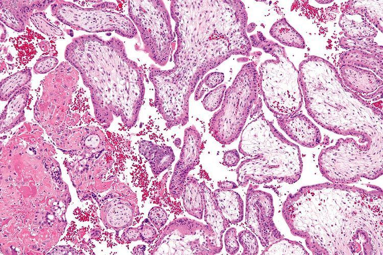
Histology
The bulk of the villi consist of connective tissues that contain blood vessels. Most of the cells in the connective tissue core of the villi are fibroblasts. Macrophages known as Hofbauer cells are also present.
Use for prenatal diagnosis
In 1983, an Italian biologist named Giuseppe Simoni discovered a new method of prenatal diagnosis using chorionic villi.
Stem cell
Chorionic villi are a rich source of stem cells. Biocell Center, a biotech company managed by Giuseppe Simoni, is studying and testing these types of stem cells. Chorionic stem cells, like amniotic stem cells, are uncontroversial multipotent stem cells.