
FMA 58279 | TA A04.4.02.013 | |
 | ||
Latin Centrum tendineum diaphragmatis | ||
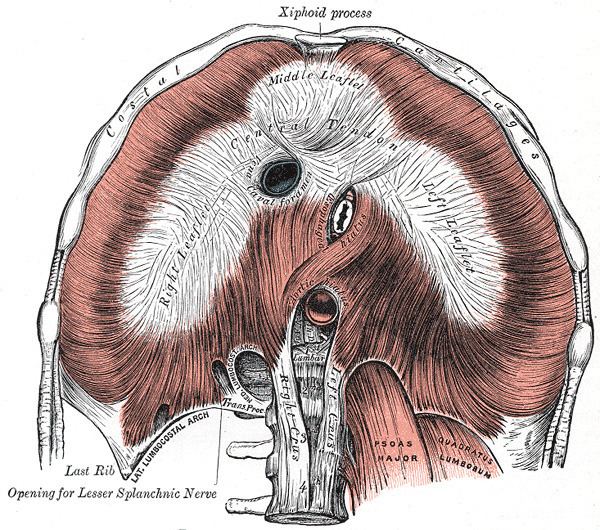
The central tendon of the diaphragm is a thin but strong aponeurosis situated slightly anterior to the vault formed by the muscle, resulting in longer posterior muscle fibers.
Contents
It is inferior to the fibrous pericardium, which fuses with the central tendon of the diaphragm via the pericardiacophrenic ligament.
The caval opening (at the level of the T8 vertebra) passes through the central tendon. This transmits the inferior vena cava and right phrenic nerve.
Structure
It is shaped somewhat like a trefoil leaf, consisting of three divisions or leaflets separated from one another by slight indentations.
The right leaflet is the largest, the middle, directed toward the xiphoid process, the next in size, and the left the smallest.
In structure the tendon is composed of several planes of fibers, which intersect one another at various angles and unite into straight or curved bundles—an arrangement which gives it additional strength.
Action during respiration
During respiration the diaphragm contracts causing the central tendon to be drawn inferiorly which partially flattens the domes. The result is an enlargement of thoracic cavity and reduction in intra-thoracic pressure. Physiologically this means that air enters the lungs and venous return to the heart is enhanced. During inspiration the central tendon retains its shape due to its tendinous nature and prevents constriction of the inferior vena cava or aorta, however the esophagus is surrounded by muscle at the esophageal hiatus and is constricted.