
 | ||
The biuret test (Piotrowski's test) is a chemical test used for detecting the presence of peptide bonds. In the presence of peptides, a copper(II) ion forms violet-colored coordination complexes in an alkaline solution. Several variants on the test have been developed, such as the BCA test and the Modified Lowry test.
Contents
The biuret reaction can be used to assess the concentration of proteins because peptide bonds occur with the same frequency per amino acid in the peptide. The intensity of the color, and hence the absorption at 540 nm, is directly proportional to the protein concentration, according to the Beer-Lambert law.
Despite its name, the reagent does not in fact contain biuret ((H2N-CO-)2NH). The test is named so because it also gives a positive reaction to the peptide-like bonds in the biuret molecule.
Procedure
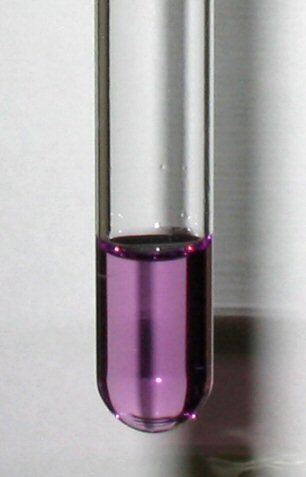
An aqueous sample is treated with an equal volume of 1% strong base (sodium or potassium hydroxide) followed by a few drops of aqueous copper(II) sulfate. If the solution turns purple, protein is present. 5–160 mg/mL can be determined. Peptides with the chain length of at least 3 amino acids are necessary for a significant, measurable colour shift with these reagents.
Biuret reagent
The Biuret reagent is made of sodium hydroxide (NaOH) and hydrated copper(II) sulfate, together with potassium sodium tartrate. Potassium sodium tartrate is added to chelate and thus stabilize the cupric ions. The reaction of the cupric ions with the nitrogen atoms involved in peptide bonds leads to the displacement of the peptide hydrogen atoms under the alkaline conditions. A tri or tetra dentate chelation with the peptide nitrogen produces the "biuret" color. This is found with dipeptides (Datta,S.P., Leberman,R., and Rabin,B.R., Trans.Farad.Soc. (1959), 55, 2141.)
The reagent is commonly used in the biuret protein assay, a colorimetric test used to determine protein concentration by UV/VIS spectroscopy at wavelength 550 nm.
High sensitivity variants of the biuret test
Two major modifications of the biuret test are commonly applied in modern colorimetric analysis of peptides: the bicinchoninic acid (BCA) assay and the Lowry assay. In these tests, the Cu+ formed during the biuret reaction reacts further with other reagents, leading to a deeper color.
In the BCA test, Cu+ forms a deep purple complex with bicinchoninic acid (BCA), which absorbs around 562 nm, producing the signature violet color. The water-soluble BCA/copper complex absorbs much more strongly than the peptide/copper complex, increasing the sensitivity of the biuret test by a factor of around 100: the BCA assay allows to detect proteins in the range of 0.0005 to 2 mg/mL). Additionally, the BCA protein assay gives the important benefit of compatibility with substances like up to 5% surfactants in protein samples.
In the Lowry protein assay Cu+ is oxidized back to Cu2+ by MoVI in Folin-Ciocalteu's reagent, which forms molybdenum blue (MoIV). Tyrosine residues in the protein also form molybdenum blue under these circumstances. In this way, proteins can be detected in concentrations between 0.005 and 2 mg/mL. Molybdenum blue in turn can bind certain organic dyes such as malachite green and Auramin O, resulting in further amplification of the signal.