
Specialty medical genetics ICD-9-CM 751.61 DiseasesDB 1400 | ICD-10 Q44.2 OMIM 210500 MedlinePlus 001145 | |
 | ||
Biliary atresia, also known as extrahepatic ductopenia and progressive obliterative cholangiopathy, is a childhood disease of the liver in which one or more bile ducts are abnormally narrow, blocked, or absent. It can be congenital or acquired. As a birth defect in newborn infants, it has an incidence of one in 10,000–15,000 live births in the United States, and a prevalence of one in 16,700 in the British Isles. Biliary atresia is most common in East Asia, with a frequency of one in 5,000.
Contents
The causes of biliary atresia are not well understood. Congenital biliary atresia has been associated with certain genes, while acquired biliary atresia is thought to be a result of an autoimmune inflammatory response, possibly due to a viral infection of the liver soon after birth. The only effective treatments are surgeries such as the Kasai procedure and liver transplantation.
Signs and symptoms
Initially, the symptoms of biliary atresia are indistinguishable from those of neonatal jaundice, a usually harmless condition commonly seen in infants. Distinctive symptoms of biliary atresia are usually evident between one and six weeks after birth. Infants and children with biliary atresia develop progressive cholestasis, a condition in which bile is unable to leave the liver and builds up inside of it. When the liver is unable to excrete bilirubin through the bile ducts in the form of bile, bilirubin begins to accumulate in the blood, causing symptoms. These symptoms include yellowing of the skin, itchiness, poor absorption of nutrients (causing delays in growth), pale stools, dark urine, and a swollen abdomen. Eventually, cirrhosis with portal hypertension will develop. If left untreated, biliary atresia can lead to liver failure. Unlike other forms of jaundice, however, biliary-atresia-related cholestasis mostly does not result in kernicterus, a form of brain damage resulting from liver dysfunction. This is because in biliary atresia, the liver, although diseased, is still able to conjugate bilirubin, and conjugated bilirubin is unable to cross the blood–brain barrier.
Pathophysiology
The cause of biliary atresia in most infants is not known and it is likely that a number of factors may play a role. Some may be due to a defect in early bile duct development (particularly those with other abnormalities) and some may arise in the perinatal period due to an external cause such as an hepatotropic virus reovirus 3 infection, congenital cytomegalovirus infection, and autoimmunity. However, experimental evidence is insufficient to confirm any of these theories.
In an Egyptian study, abnormally high levels of aflatoxin B1 and to a lesser extent aflatoxin B2 was found in liver tissue and blood of all neonates suffering from biliary atresia. Aflatoxins may cause extensive damage to the hepatocytes leading to hepatitis and damage to bile ducts causing inflammation, adhesions and final obstruction of bile ducts. The affected neonates have a genetic detoxification defect that does not allow them to detoxify these aflatoxins timely or effectively. The babies have homozygous deficiency of glutathione S transferase (GST) M1. The aflatoxin damaged liver cells and bile duct cells are removed by neutrophil elastase and by involvement of immune system mediators such as CCL-2 or MCP-1, tumor necrosis factor (TNF), interleukin-6 (IL-6), TGF-beta, endothelin (ET), and nitric oxide (NO). Among these, TGF-beta is the most important pro-fibrogenic cytokine that can be seen in progressive cirrhosis.
The cascade of immune involvement to remove damaged hepatocytes and cholangiocytes ushers regeneration. Yet in infants with biliary atresia regeneration is defective, and results in cirrhosis, as these infants have disrupted p53 and disrupted GSTPi. p53 and GSTPi are responsible for DNA fidelity at regeneration. Hence, these infants get accelerated cirrhosis and march to portal hypertension.
Progressive cirrhosis, is associated with signs and symptoms of portal hypertension, such as esophagogastric varix bleeding, hypersplenism, hepatorenal syndrome, and hepatopulmonary syndrome.
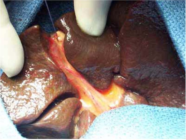
There are three main types of extra-hepatic biliary atresia:
In approximately 10% of cases, anomalies associated with biliary atresia include heart lesions, polysplenia, situs inversus, absent venae cavae, and a preduodenal portal vein.
Genetics
An association between biliary atresia and the ADD3 gene was first detected in Chinese populations through a Genome-wide association study, and was confirmed in Thai Asians and Caucasians. A possible association with deletion of the gene GPC1, which encodes a glypican 1-a heparan sulfate proteoglycan, has been reported. This gene is located on the long arm of chromosome 2 (2q37) and is involved in the regulation of inflammation and the Hedgehog gene.
Neonates with biliary atresia were found to have null GSTM1 genotype while all their moms were heterozygous for GSTM1. Thus these infants are protected intrauterine by their maternal detoxification system, yet once born they cannot handle the detoxification of aflatoxin load.
Toxins
Some cases of biliary atresia may result from exposure to aflatoxin B1, and to a lesser extent aflatoxin B2 during late pregnancy. Intact maternal detoxification protects baby during intrauterine life, yet after delivery the baby struggles with the aflatoxin in its blood and liver. Moreover, the baby feeds aflatoxin M1 from its mom, as aflatoxin M1 is the detoxification product of aflatoxin B1. It is a milder toxin that causes cholangitis in the baby.
There are isolated examples of biliary atresia in animals. For instance, lambs born to sheep grazing on land contaminated with a weed (Red Crumbweed) developed biliary atresia at certain times. The plants were later found to contain a toxin, now called biliatresone Studies are ongoing to determine whether there is a link between human cases of biliary atresia and toxins such as biliatresone. There are some indications that a metabolite of certain human gut bacteria may be similar to biliatresone.
Diagnosis
Diagnosis is made by an assessment of symptoms, physical exam, and medical history, in conjunction with blood tests, a liver biopsy, and imaging. Diagnosis is often made following investigation of prolonged jaundice that is resistant to phototherapy and/or exchange transfusions, with abnormalities in liver enzyme tests. Ultrasound or other forms of imaging can confirm the diagnosis. Further testing may include radioactive scans of the liver and a liver biopsy.
Treatment
Most (>95%) infants with biliary atresia will undergo an operation designed to retain and salvage the native liver, restore bile flow and reduce the level of jaundice. This is known as the Kasai procedure (after Morio Kasai, the Japanese surgeon who first developed the technique) or hepatoportoenterostomy. Although the procedure is not thought of as curative, it may relieve jaundice, and stop liver fibrosis allowing normal growth and development. Published series from Japan, North America and the UK show that bilirubin levels will fall to normal values in about 50-55% of infants allowing 40-50% to retain their own liver to reach the age of 5 and 10 years (and beyond). Liver transplantation is an option for those children whose liver function and symptoms fail to respond to a Kasai operation.
Recent large-scale studies by Davenport et al. (Annals of Surgery, 2008) show that the age of the patient is not an absolute clinical factor affecting prognosis. The influence of age differs according to the disease etiology—i.e., whether biliary atresia is isolated, cystic (CBA), or accompanied by splenic malformation (BASM).
It is widely accepted that corticosteroid treatment after a Kasai operation, with or without choleretics and antibiotics, has a beneficial effect on postoperative bile flow and can clear jaundice, but the dosing and duration of the ideal steroid protocol are controversial. Furthermore, it has been observed in many retrospective longitudinal studies that corticosteroid treatment does not prolong survival of the native liver or transplant-free survival. Davenport et al. also showed (Hepatology 2007) that short-term, low-dose steroid therapy following a Kasai operation had no effect on the mid- or long-term prognosis of biliary atresia patients.
Epidemiology
Biliary atresia seems to affect females slightly more often than males, and Asians and African Americans more often than Caucasians. It is common for only one child in a pair of twins or within the same family to have the condition. There seems to be no link to medications or immunizations given immediately before or during pregnancy. Diabetes during pregnancy particularly during the first trimester seems to predispose to a number of distinct congenital abnormalities in the infant such as sacral agenesis and the syndromic form of biliary atresia.