
Specialty neurology ICD-9-CM 341.1 MeSH D002549 | ICD-10 G37.5 DiseasesDB 11849 | |
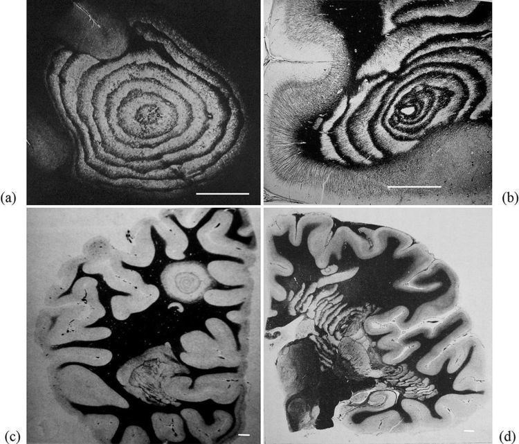 | ||
Balo concentric sclerosis is a disease in which the white matter of the brain appears damaged in concentric layers, leaving the axis cylinder intact. It was described by Joszef Balo who initially named it "leuko-encephalitis periaxialis concentrica" from the previous definition, and it is currently considered one of the borderline forms of multiple sclerosis.
Contents
- Pathophysiology
- Lesions under MRI
- Models
- Spectrum
- Paediatric cases
- Lesions in autopsy and biopsy
- Epidemiology
- History
- References
Balo concentric sclerosis is a demyelinating disease similar to standard multiple sclerosis, but with the particularity that the demyelinated tissues form concentric layers. Scientists used to believe that the prognosis was similar to Marburg multiple sclerosis, but now they know that patients can survive, or even have spontaneous remission and asymptomatic cases.
It is also common that the clinical course is primary progressive, but a relapsing-remitting course has been reported. It seems that the course gets better with prednisone therapy, although evidence of this is anecdotal and such conclusions are difficult to accept given that there are cases where patients spontaneously recover whether the patient was on steroid therapy or not.
Pathophysiology
The lesions of the Balo sclerosis belong to the MS lesion pattern III (distal oligodendrogliopathy). Balo concentric sclerosis is now believed to be a variant of pattern III multiple sclerosis.
According with Dr. Lucchinetti investigations, in Balo's concentric sclerosis, the rings may be caused by a physiological hypoxia (similar to that caused by some toxins or viruses) in the lesion, which is in turn countered by expression of stress proteins at the border. This expression and counter-expression forms rings of preserved tissue within the lesion and rings of demyelinated tissue just beyond where the previous attack had induced the protective stress proteins. Hence, subsequent attacks form concentric rings.
Some other researchers maintain that, as in pattern III MS, the mitochondrial respiratory chain complex IV activity is reduced and this could be the culprit of glutamate-mediated axonal injury.
Other two propossed mechanisms are the presence of granulation tissue in old lesions, which is rich in capillaries and could act as an energy reservoir, and the small adherent bands of myelinated tissue. These two mechanisms are proposed based in the assumption of a hypoxic causation principle
Ultimately, this expanding lesion causes the progressive picture typically seen. However, in some patients, the pathology underlying the disease appears to burn out and hence the disease may halt, hence the patients who spontaneously recover. The mechanisms triggering attacks and recovery remain uncertain.
Nevertheless, this model is questioned by recent reports that found astrocyte damage, similar to the one found in aquaporin-seropositive neuromyelitis optica. Though no anti-NMO antibodies have been found, the damage is similar, pointing to problems in the water channel of the astrocytes
It presents three clinical subtypes: Monophasic, relapsing-remitting and primary rapidly progressive. CSF is either normal or shows mild mononuclear inflammatory reaction. Oligoclonal bands are present only in few cases
Lesions under MRI
The features of the MRI and the characteristics of the lesion can be correlated when a biopsy has been taken, providing a way to standarize the future MRI diagnosis
Balo concentric sclerosis lesions can be distinguished from normal lesions on MRI showing alternative hypotense and hypertense layers
Balo concentric lesions can be viewed using the myelin water imaging techniques. This is a special MRI sequence that shows the myelin's percentage of water content.
Pattern III lesions, including Balo lesions, have a specific initiation pattern under MRI (MRILIP) consisting in showing Gadolinium enhancement before FLAIR MRI appearance.
Models
A mathematical model for concentric sclerosis has been proposed. Authors review the previous pathogenic theories, discuss the link between concentric sclerosis and Liesegang's periodic precipitation phenomenon and propose a new mechanism based on self-organization.
Spectrum
Balo lesions have been reported alone, but also associated to standard multiple sclerosis, neuromyelitis optica, CADASIL and progressive multifocal leukoencephalopathy
Paediatric cases
Balo concentric sclerosis in children has been reported to behave different from adults
Lesions in autopsy and biopsy
A report comparing 1H-magnetic resonance spectroscopy, magnetization transfer and diffusion tensor imaging with histopathology in a patient with Balo's concentric sclerosis, found that inflammation was traced by fractional anisotropy and increased lactate. In contrast, magnetization transfer ratio and the diffusion coefficient show a loss of tissue in the rings of the lesion.
Epidemiology
The disease is more common in Chinese and Filipino populations (both Asiatic) than in caucasoids.
History
Though the disease carries the name of Joszef Balo, it was first described by Otto Marburg in 1906 Later, in 1928, Joszef Balo studied the encephalitis periaxialis concentrica in a Hungarian patient, showing also demyelination of the peripheral nervous system.