
Latin auricula FMA 56580 | TA A15.3.01.002 | |
 | ||
Artery posterior auricular, anterior auricular Lymph To Pre & Post Auricular Nodes, Nodes of Parotid and Cervical Chains | ||
The auricle or auricula is the visible part of the ear that resides outside of the head. It is also called the pinna (Latin for wing / fin, plural pinnae), a term that is used more in zootomy.
Contents
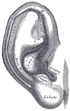
Structure
The diagram shows the shape and location of most of these components:
Development
The developing auricle is first noticeable around the sixth week of gestation in the human fetus, developing from the auricular hillocks, which are derived from the first and second pharyngeal arches. These hillocks develop into the folds of the auricle and gradually shift upwards and backwards to their final position on the head. En route accessory auricles (also known as preauricular tags) may be left behind. The first three hillocks are derived from the 1st branchial arch and form the tragus, crus of the helix, and helix, respectively. Cutaneous sensation to these areas is via the trigeminal nerve, the attendant nerve of the 1st branchial arch. The final three hillocks are derived from the second branchial arch and form the antihelix, antitragus, and lobule, respectively. These portions of the ear are supplied by the cervical plexus and a small portion by the facial nerve. This explains why vesicles are classically seen on the auricle in herpes infections of the facial nerve (Ramsay Hunt syndrome type II).
Function
The auricle functions to collect sound and transform it to directional and other information. The auricle collects sound and acting as a funnel, amplifies the sound and directs it to the auditory canal. The filtering effect of the human pinna preferentially selects sounds in the frequency range of human speech.
Amplification
Amplification of sound by the pinna, tympanic membrane and middle ear causes an increase in level of about 10 to 15 dB in a frequency range of 1.5 kHz to 7 kHz. This amplification is an important factor in inner ear trauma resulting from elevated sound levels.
Notch of pinna
Due to its anatomy, the pinna largely eliminates a small segment of the frequency spectrum; this band is called the pinna notch. The pinna works differently for low and high frequency sounds. For low frequencies, it behaves similarly to a reflector dish, directing sounds toward the ear canal. For high frequencies, however, its value is thought to be more sophisticated. While some of the sounds that enter the ear travel directly to the canal, others reflect off the contours of the pinna first: these enter the ear canal after a very slight delay. This delay causes phase cancellation, virtually eliminating the frequency component whose wave period is twice the delay period. Neighboring frequencies also drop significantly. In the affected frequency band – the pinna notch – the pinna creates a band-stop or notch filtering effect. This filter typically affects sounds around 10 kHz, though it can affect any frequencies from 6 – 16 kHz. It also is directionally dependent, affecting sounds coming from above more than those coming from straight ahead. This aids in vertical sound localization.
Other animals
In animals the function of the pinna is to collect sound, and perform spectral transformations to incoming sounds which enable the process of vertical localization to take place. It collects sound by acting as a funnel, amplifying the sound and directing it to the auditory canal. While reflecting from the pinna, sound also goes through a filtering process, as well as frequency dependent amplitude modulation which adds directional information to the sound (see sound localization, vertical sound localization, head-related transfer function, pinna notch). In various species, the pinna can also signal mood and radiate heat.
Clinical significance
There are various visible ear abnormalities: