
 | ||
Apoptotic DNA fragmentation is a key feature of apoptosis, a type of programmed cell death. Apoptosis is characterized by the activation of endogenous endonucleases with subsequent cleavage of chromatin DNA into internucleosomal fragments of roughly 180 base pairs (bp) and multiples thereof (360, 540 etc.). This effect can be used to detect apoptosis, for example via the DNA laddering assay, the TUNEL assay, or the Nicoletti assay.
Contents
Mechanism
The enzyme responsible for apoptotic DNA fragmentation is the Caspase-Activated DNase (CAD). CAD is normally inhibited by another protein, the Inhibitor of Caspase Activated DNase (ICAD). During apoptosis, the apoptotic effector caspase, caspase 3, cleaves ICAD and thus causes CAD to become activated.
CAD cleaves DNA at internucleosomal linker sites between nucleosomes, protein-containing structures that occur in chromatin at ~180-bp intervals. This is because the DNA is normally tightly wrapped around histones, the core proteins of the nucleosomes. The linker sites are the only parts of the DNA strand that are exposed and thus accessible to CAD.
Degradation of nuclear DNA into nucleosomal units is one of the hallmarks of apoptotic cell death. It occurs in response to various apoptotic stimuli in a wide variety of cell types. Molecular characterization of this process identified a specific DNase (CAD, caspase-activated DNase) that cleaves chromosomal DNA in a caspase-dependent manner. CAD is synthesized with the help of ICAD (inhibitor of CAD), which works as a specific chaperone for CAD and is found complexed with ICAD in proliferating cells. When cells are induced to undergo apoptosis, caspase 3 cleaves ICAD to dissociate the CAD:ICAD complex, allowing CAD to cleave chromosomal DNA. Cells that lack ICAD or that express caspase-resistant mutant ICAD thus do not show DNA fragmentation during apoptosis, although they do exhibit some other features of apoptosis and die.
DNA fragmentation is a secondary consequence, rather than an integral cause, of apoptosis. Endonuclease involved might be similar to DNase I, a potential indication that DNA fragmentation might occur after the release of enzymes from the plasma membrane lysis, an event that would potentially occur only after the final lytic event in the apoptotic sequence. More recently, data have shown that specific proteases residing in the cytoplasm mediate the terminal events of apoptosis, including those of nuclear morphology. Even so, the detection of DNA fragmentation and the presence of single strand ends of DNA have continued to be used in many studies to detect apoptotic cells, particularly in intact tissues, though necrosis also produces single-strand DNA ends in cell nuclei. Therefore, the interpretation of these in situ assays of DNA fragmentation—in situ nick translation (ISNT), TUNEL assay—must be carefully assessed together with morphological features of apoptotic cells.
Even though much work has been performed on the analysis of apoptotic events, little information is available to link the timing of morphological features at the cell surface and in the nucleus to the biochemical degradation of DNA in the same cells. Apoptosis can be initiated by a myriad of different mechanisms in different cell types, and the kinetics of these events vary widely, from only a few minutes to several days depending on the cell system.
Historical background
The discovery of the internucleosomal fragmentation of genomic DNA to regular repeating oligonucleosomal fragments generated by Ca/Mg-dependent endonuclease is accepted as one of the best-characterized biochemical markers of apoptosis (programmed cell death).
In 1970, Williamson described that cytoplasmic DNA isolated from mouse liver cells after culture was characterized by DNA fragments with a molecular weight consisting of multiples of 135 kDa. This finding was consistent with the hypothesis that these DNA fragments were a specific degradation product of nuclear DNA. In 1972, Kerr, Wyllie, and Currie coined the term apoptosis and distinguished this type of cell death from necrosis based on morphological features. In 1973, Hewish and Burgoyne, during the study of subchromatin structure, found that chromatin is accessible to the Ca++/Mg++ endonuclease, resulting in the formation of a digestion product with a regular series of molecular weight similar to the one previously described by Williamson (1970). In 1974, Williams, Little, and Shipley, using cells exposed to widely differing types of trauma, found that during cell death, degraded DNA in "every case had a modal value of between 10(x6) and 10(x7) Dalton and cellular metabolism is required to produce degradation of DNA". However, this observation was without indication of "whether the incision attack on the DNA molecule was a random or rather at a particular site, that have structural or functional meaning". In 1976, Scalka, Matyasova, and Cejkova described internucleosomal fragmentation of irradiated lymphoid chromatin DNA in vivo.
Six years passed from 1972 to 1978/1980 until the discovery and evaluation of internucleosomal fragmentation of DNA during apoptotic cell death as a hallmark of apoptosis. Since 1972 (Kerr, Wyllie, and Currie), it is accepted that glucocorticoid-induced death of lymphocytes is a form of apoptosis. In 1978, Zakharyan and Pogosyan presented a paper revealing that glucocorticoid-induced DNA degradation in rat lymphoid tissue, thymus, and spleen occurred in a specific pattern producing fragments of DNA that were electrophoretically similar to those observed after treatment of chromatin with microccoccal nuclease, which indicated internucleosomal cleavage pattern of DNA degradation occurred during apoptosis. Thus, the first link between programmed cell death/apoptosis and internucleosomal fragmentation of chromatin DNA was discovered and soon became as a specific feature of apoptosis.
In 1980, Wyllie reported additional evidence for an internucleosomal DNA cleavage pattern as a specific feature of glucocorticoid-treated thymocytes undergoing apoptosis. The internucleosomal DNA cleavage pattern was observed as a specific feature of apoptosis in 1978/1980 and has become a recognised hallmark of programmed cell death since then.
Detection assays
There are many methods to assess the DNA fragmentation caused by apoptosis and also many commercial kits are available. Some new methods do quantification assays for DNA Fragmentation by flow cytometry and fluorescent assays.
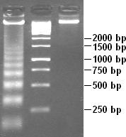
Apoptotic DNA fragmentation is often analyzed using agarose gel electrophoresis to demonstrate a "ladder" pattern at ~180-BP intervals. Necrosis, on the other hand, is usually characterized by random DNA fragmentation which forms a "smear" on agarose gels.