
Entrez 9131 OMIM 300169 | Alt. symbols PDCD8 HUGO 8768 RefSeq NM_004208 | |
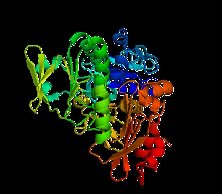 | ||
Apoptosis inducing factor is a flavoprotein.
Contents
Apoptosis inducing factor is involved in initiating a caspase-independent pathway of apoptosis (positive intrinsic regulator of apoptosis) by causing DNA fragmentation and chromatin condensation. It also acts as an NADH oxidase. Another AIF function is to regulate the permeability of the mitochondrial membrane upon apoptosis. Normally it is found behind the outer membrane of the mitochondria and is therefore secluded from the nucleus. However, when the mitochondrion is damaged, it moves to the cytosol and to the nucleus. Inactivation of AIF leads to resistance of embryonic stem cells to death following the withdrawal of growth factors indicating that it is involved in apoptosis.
Function
Apoptosis Inducing Factor (AIF) is a protein that triggers chromatin condensation and DNA fragmentation in a cell in order to induce programmed cell death. The mitochondrial AIF protein was found to be a caspase-independent death effector that can allow independent nuclei to undergo apoptotic changes. The process triggering apoptosis starts when the mitochondria releases AIF, which exits through the mitochondrial membrane, enters the cytosol, and finally ends up in the cell nucleus where it signals the cell to condense its chromosomes and fragment its DNA molecules in order to prepare for cell death. Recently, researchers have discovered that AIF's activity is dependent upon the type of cell, the apoptotic insult, and its DNA-binding ability. AIF also plays a significant role in the mitochondrial respiratory chain and metabolic redox reactions.
Synthesis
The AIF protein is located across 16 exons on the X chromosome in humans. AIF1 (the most abundant type of AIF) is translated in the cytosol and recruited to the mitochondrial membrane and intermembrane space by its N-terminal mitochondrial localization signal (MLS). Inside the mitochondria, AIF folds into its functional configuration with the help of the cofactor flavin adenine dinucleotide (FAD).
A protein called Scythe (BAT3), which is used to regulate organogenesis, can increase the AIF lifetime in the cell. As a result, decreased amounts of Scythe lead to a quicker fragmentation of AIF. The X-linked inhibitor of apoptosis (XIAP) has the power to influence the half-life of AIF along with Scythe. Together, the two do not affect the AIF attached to the inner mitochondrial membrane, however they influence the stability of AIF once it exits the mitochondria.
Role in mitochondria
It was thought that if a recombinant version of AIF lacked the first N-terminal 120 amino acids of the protein, then AIF would function as an NADH and NADPH oxidase. However, it was instead discovered that recombinant AIF that do not have the last 100 N-terminal amino acids have limited NADP and NADPH oxidase activity. Therefore, researchers concluded that the AIF N-terminus may function in interactions with other proteins or control AIF redox reactions and substrate specificity.
Mutations of AIF due to deletions have stimulated the creation of the mouse model of complex I deficiency. Complex I deficiency is the reason behind over thirty percent of human mitochondrial diseases. For example, complex I mitochondriopathies mostly affect infants by causing symptoms such as seizures, blindness, deafness, etc. These AIF-deficient mouse models are important for fixing complex I deficiencies. The identification of AIF-interacting proteins in the inner mitochondrial membrane and intermembrane space will help researchers identify the mechanism of the signaling pathway that monitors the function of AIF in the mitochondria.
Isozymes
Human genes encoding apoptosis inducing factor isozymes include: