
TA A01.2.02.002 | FMA 57777 | |
 | ||
Latin Trigonum cervicale anteriusTrigonum colli anteriusRegio cervicalis anterior | ||
The anterior triangle is a region of the neck.
Contents
Structure
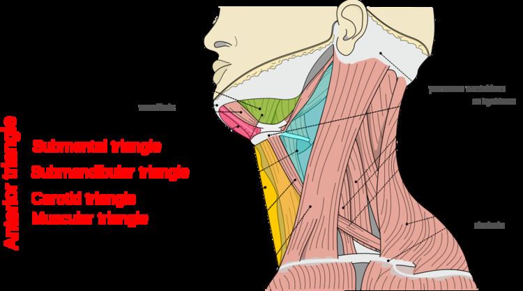
The triangle is inverted with its apex inferior to its base which is under the chin.
Investing fascia covers the roof of the triangle while visceral fascia covers the floor.
Anatomy
Muscles:
Nerve supply
2 Bellies of Digastric
Stylohyoid: by the facial nerve, by a branch from that to the posterior belly of digastric.
Mylohyoid: by its own nerve, a branch of the inferior alveolar ( from the mandibular division of trigemminal nerve), which arises just before the parent nerve enters the mandibular foramen, pierces the sphenomandibular ligament, and runs forward on the inferior surface of the mylohyoid, supplying it and the anterior belly of the digastric.
Geniohyoid: by a branch from the hypoglossal nerve consisting of fibres from the C1 nerve.
Sternohyoid, Omohyoid, Sternothyroid are supplied by Ansa cervicalis.
Thyrohyoid: by a branch of hypoglossal nerve but the fibres are all 'hitch-hiking' from C1.
Development
Divisions
This space is subdivided into four smaller triangles by the Digastricus above, and the superior belly of the Omohyoideus.
These smaller triangles are named: