
Group Group I (dsDNA) Scientific name Whispovirus: Rank Genus | Family Nimaviridae Higher classification Nimaviridae | |
 | ||
Similar Penaeus, Infectious hypodermal and hem, Freshwater white spot disease, Whiteleg shrimp, Giant tiger prawn | ||
White spot syndrome virus is the lone virus (and type species) of the genus Whispovirus (white spot), which is the only genus in the family Nimaviridae. It is responsible for causing white spot syndrome in a wide range of crustacean hosts. White spot syndrome (WSS) is a viral infection of penaeid shrimp. The disease is highly lethal and contagious, killing shrimps quickly. Outbreaks of this disease have wiped out within a few days the entire populations of many shrimp farms throughout the world.
Contents
- Bt white spot syndrome virus tumama sa shrimp farms sa sarangani at gensan
- History
- Virology
- Taxonomy
- Virion Structure
- Genome
- Life Cycle
- Clinical
- Pathology
- Diagnosis
- Treatment
- Prevention
- References
The disease is caused by a family of related viruses subsumed as the Whitespot Syndrome Baculovirus complex (WSSV) and the disease caused by them as white spot syndrome (WSS).
Bt white spot syndrome virus tumama sa shrimp farms sa sarangani at gensan
History
The first reported epidemic due to this virus is from Taiwan in 1992; however, reports of losses due to white spot disease came from China in 1993, where it led to a virtual collapse of the shrimp farming industry. This was followed by outbreaks in Japan and Korea in the same year, Thailand, India and Malaysia in 1994, and by 1996 it had severely affected East Asia and South Asia. In late 1995, it was reported in the United States, 1998 in Central and South America, 1999 in Mexico' in 2000 in the Philippines, and in 2011 in Saudi Arabia. The virus was detected in Queensland, Australia in November 2016. Australia was believed to be the last shrimp-growing region free from the virus.
Virology
White spot syndrome virus is a species of virus in the genus Whispovirus, in the family Nimaviridae. It is the only species in this family, and so serves as the type species.
Taxonomy
White spot syndrome virus is a dsDNA virus, which places it in Group I of the Baltimore classification system.
Group: dsDNA
Virion Structure
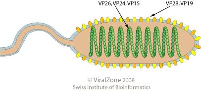
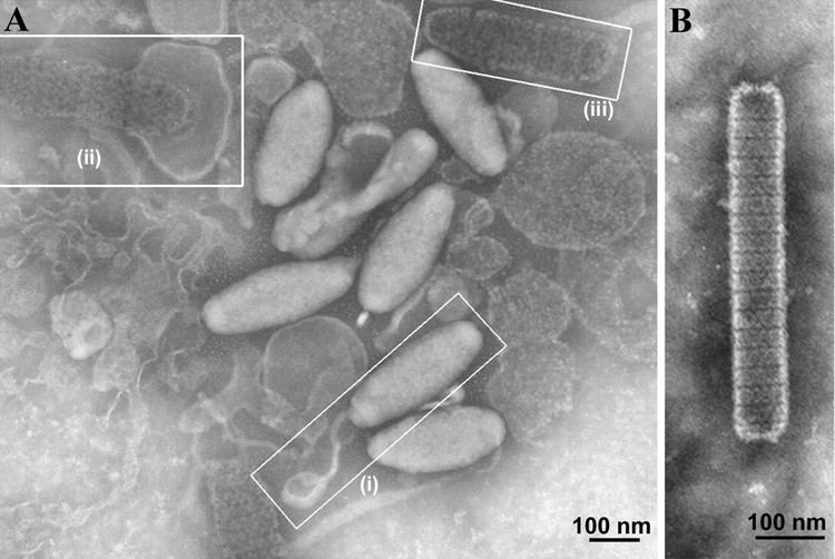
WSSV is a rod-shaped, double-stranded, DNA virus, and the size of the enveloped viral particles have been reported to be 240–380 nm long and 70–159 nm in diameter and nucleocapsid core is 120–205 nm long and 95–165 nm in diameter. The virus has an outer lipid bilayer membrane envelope, sometimes with a tail like appendage at one end of the virion. The nucleocapsid consists of 15 conspicuous vertical helices located along the long axis, each helix has two parallel striations, composed of 14 globular capsomers, each of which is 8 nm in diameter.
Genome
The complete DNA sequence of WSSV genome has been assembled into a circular sequence of 292,967 bp. It encodes 531 putative open reading frames.
One of the proteins – WSSV449 – has some similarity to host protein Tube and can function like Tube by activating the NF-κB pathway.
Life Cycle
Viral replication is nuclear. DNA-templated transcription is the method of transcription. The virus infects an unusually wide host range of crustaceans. Transmission of the virus is mainly through oral ingestion and water-borne routes in farms (horizontal transmission) and vertical transmission (from infected mother prawns) in the case of shrimp hatcheries. The virus is present in the wild stocks of shrimp, especially in the coastal waters adjacent to shrimp farming regions in Asian countries, but mass mortalities of wild shrimps are not yet to be observed.
Clinical
The virus has a wide host range. While shrimp can survive with the virus for extended periods of time, factors like stress can cause the outbreak of WSS. The disease is highly virulent and leads to mortality rates of 100% within days in the case of cultured penaeid shrimps. Most of the cultured penaeid shrimps (Penaeus monodon, Marsupenaeus japonicus, Litopenaeus vannamei, and Fenneropenaeus indicus) are natural hosts of the virus. Several non-penaeid shrimps were also found to be severely infected during experimental challenges. Many crustaceans like crabs (Scylla spp., Portunus spp.), spiny lobsters (Panulirus spp.), crayfish (Astacus spp., Cherax spp.) and freshwater shrimp (Macrobrachium spp.) are reported to be infected with variable severities depending on the life stage of the host and presence of external stressors (temperature, salinity, bacterial diseases, pollutants).
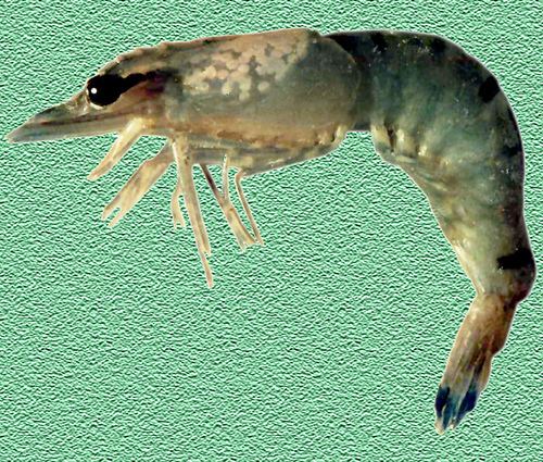
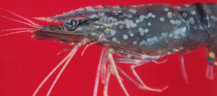
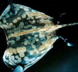
Clinical signs of WSS include a sudden reduction in food consumption, lethargy, loose cuticle and often reddish discolouration, and the presence of white spots of 0.5 to 2.0 mm in diameter on the inside surface of the carapace, appendages and cuticle over the abdominal segments.
Pathology
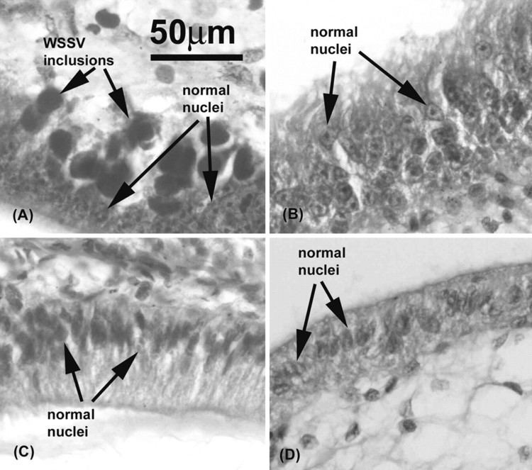
In the host, WSSV infects a wide variety of cells from ectodermal and mesodermal origin. Histological changes are seen in the gill epithelium, antennal gland, haematopoeitic tissue, nervous tissue, connective tissue and intestinal epithelial tissue. Infected cells have prominent intranuclear occlusions that initially stain eosinophilic, but become basophilic with age; hypertrophied nuclei with chromatin margination; and cytoplasmic clearing. Pathogenesis involves widespread tissue necrosis and disintegration.
White spots on the shell of infected shrimp under scanning electron microscope appear as large, dome-shaped spots on the carapace measuring 0.3 to 3 mm in diameter. Smaller white spots of 0.02 to 0.1 mm appear as linked spheres on the cuticle surface. Chemical composition of the spots is similar to the carapace, calcium forming 80–90% of the total material and it is suggested to have derived from abnormalities of the cuticular epidermis.
A number of biochemical changes have been reported after infection with this virus: glucose consumption and plasma lactate concentration increase, glucose 6 phosphate dehydrogenase activity increases and triglyceride concentration decreases. The voltage dependent anion channel of the mitochondrion is also up regulated.
Diagnosis
Infection with WSSV differs from other described penaeid infections Yellowhead virus (YHV) and Infectious Hypodermal and Hematopoietic Necrosis virus (IHHNV) in the described histological findings as YHV has a reduced tissue specificity, infecting only the intestinal epithelial tissues and IHHNV causes intranuclear occlusions that stain eosinophillic but do not change over the course of the infection.
Rapid and specific diagnosis of the virus can be accomplished using nested or quantitative PCR.
Treatment
There are no available treatments for WSS.
Prevention
A large number of disinfectants are widely used in shrimp farms and hatcheries to prevent an outbreak. Stocking of uninfected shrimp seeds and rearing them away from environmental stressors with extreme care to prevent contamination are useful management measures. Site selection may be one of the most crucial in preventing White Spot Disease. A study showed that shrimps farmed in areas with relatively low temperature fluctuations and at water temperatures greater than 29°C had increased resistance to WSSV.