
NeuroNames ancil-252 Dorlands/Elsevier a_59/12151778 | NeuroLex ID Wernicke's area FMA 242178 | |
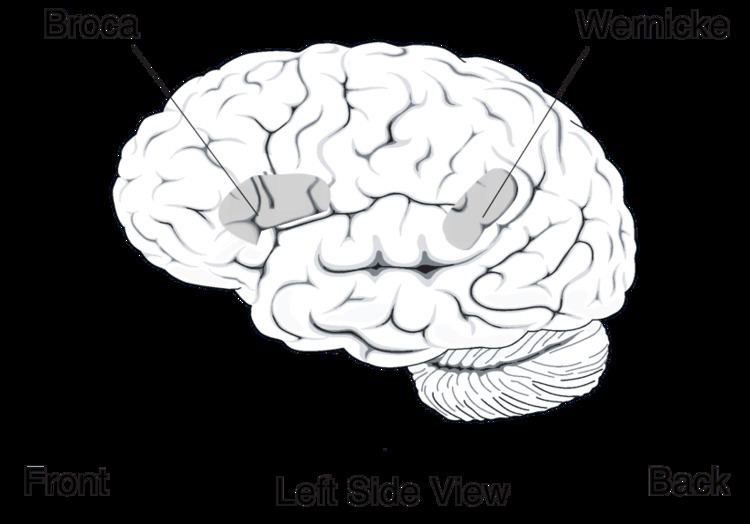 | ||
Wernicke's area (/ˈvɛərnᵻkə/ or /ˈvɛərnᵻki/; [ˈvɛʁnɪkə]), also called Wernicke's speech area, is one of the two parts of the cerebral cortex linked, since the late nineteenth century, to speech (the other is Broca's area). It is involved in the comprehension or understanding of written and spoken language (in contrast to Broca's area that is involved in the production of language). It is traditionally thought to be in Brodmann area 22, which is located in the posterior section of the superior temporal gyrus (STG) in the dominant cerebral hemisphere (which is the left hemisphere in about 95% of right handed individuals and 60% of left handed individuals). Damage caused to Wernicke's area results in receptive, fluent aphasia. This means that the person with aphasia will be able to fluently connect words, but the phrases will lack meaning. This is unlike non-fluent aphasia, in which the person will use meaningful words, but in a non-fluent, telegraphic manner.
Contents
Structure
Wernicke's area is classically located in the posterior section of the superior temporal gyrus (STG) in the (most commonly) left cerebral hemisphere. This area encircles the auditory cortex on the lateral sulcus (the part of the brain where the temporal lobe and parietal lobe meet). This area is neuroanatomically described as the posterior part of Brodmann area 22.
However, there is an absence of consistent definitions as to the location. Some identify it with the unimodal auditory association in the superior temporal gyrus anterior to the primary auditory cortex (the anterior part of BA 22). This is the site most consistently implicated in auditory word recognition by functional brain imaging experiments. Others include also adjacent parts of the heteromodal cortex in BA 39 and BA40 in the parietal lobe.
While previously thought to connect Wernicke's area and Broca's area, new research demonstrates that the arcuate fasciculus instead connects to posterior receptive areas with premotor/motor areas, and not to Broca's area. Consistent with the word recognition site identified in brain imaging, the uncinate fasciculus connects anterior superior temporal regions with Broca's area.
Right homologous area
Research using Transcranial magnetic stimulation suggests that the area corresponding to the Wernicke’s area in the non-dominant cerebral hemisphere has a role in processing and resolution of subordinate meanings of ambiguous words—such as ‘‘river’’ when given the ambiguous word "bank." In contrast, the Wernicke's area in the dominant hemisphere processes dominant word meanings (‘‘teller’’ given ‘‘bank’’).
Modern views
Neuroimaging suggests the functions earlier attributed to Wernicke's area occur more broadly in the temporal lobe and indeed happen also in Broca's area.
Support for a broad range of speech processing areas was furthered by a recent study done at University of Rochester in which American Sign Language native speakers were subject to MRIs while interpreting sentences that identified a relationship using either syntax (relationship is determined by the word order) or inflection (relationship is determined by physical motion of "moving hands through space or signing on one side of the body"). Distinct areas of the brain were activated with the frontal cortex (associated with ability to put information into sequences) being more active in the syntax condition and the temporal lobes (associated with dividing information into its constituent parts) being more active in the inflection condition. However, these areas are not mutually exclusive and show a large amount of overlap. These findings imply that while speech processing is a very complex process, the brain may be using fairly basic, preexisting computational methods.
Aphasia
Wernicke's area is named after Carl Wernicke, a German neurologist and psychiatrist who, in 1874, hypothesized a link between the left posterior section of the superior temporal gyrus and the reflexive mimicking of words and their syllables that associated the sensory and motor images of spoken words. He did this on the basis of the location of brain injuries that caused aphasia. Receptive aphasia in which such abilities are preserved is also known as Wernicke's aphasia. In this condition there is a major impairment of language comprehension, while speech retains a natural-sounding rhythm and a relatively normal syntax. Language as a result is largely meaningless (a condition sometimes called fluent or jargon aphasia).
While neuroimaging and lesion evidence generally support the idea that malfunction of or damage to Wernicke's area is common in people with receptive aphasia, this is not always so. Some people may use the right hemisphere for language, and isolated damage of Wernicke's area cortex (sparing white matter and other areas) may not cause severe receptive aphasia. Even when patients with Wernicke's area lesions have comprehension deficits, these are usually not restricted to language processing alone. For example, one study found that patients with posterior lesions also had trouble understanding nonverbal sounds like animal and machine noises. In fact, for Wernicke's area, the impairments in nonverbal sounds were statistically stronger than for verbal sounds.