
 | ||
Latin Extremitas proximalis tibiae | ||
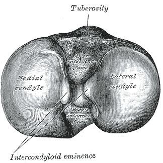
The upper extremity of the tibia or proximal extremity is large, and expanded into two eminences, the medial condyle and lateral condyle.
Contents
Facets
The superior articular surface presents two smooth articular facets.
The central portions of these facets articulate with the condyles of the femur, while their peripheral portions support the menisci of the knee joint, which here intervene between the two bones.
Intercondyloid eminence
Between the articular facets in the intercondylar area, but nearer the posterior than the anterior aspect of the bone, is the intercondyloid eminence (spine of tibia), surmounted on either side by a prominent tubercle, on to the sides of which the articular facets are prolonged; in front of and behind the intercondyloid eminence are rough depressions for the attachment of the anterior and posterior cruciate ligaments and the menisci.
Surfaces
The anterior surfaces of the condyles are continuous with one another, forming a large somewhat flattened area; this area is triangular, broad above, and perforated by large vascular foramina; narrow below where it ends in a large oblong elevation, the tuberosity of the tibia, which gives attachment to the patellar ligament; a bursa intervenes between the deep surface of the ligament and the part of the bone immediately above the tuberosity.
Posteriorly, the condyles are separated from each other by a shallow depression, the posterior intercondyloid fossa, which gives attachment to part of the posterior cruciate ligament of the knee-joint. The medial condyle presents posteriorly a deep transverse groove, for the insertion of the tendon of the semimembranosus.
Its medial surface is convex, rough, and prominent; it gives attachment to the medial collateral ligament.
The lateral condyle presents posteriorly a flat articular facet, nearly circular in form, directed downward, backward, and lateralward, for articulation with the head of the fibula. Its lateral surface is convex, rough, and prominent in front: on it is an eminence, situated on a level with the upper border of the tuberosity and at the junction of its anterior and lateral surfaces, for the attachment of the iliotibial band. Just below this a part of the extensor digitorum longus takes origin and a slip from the tendon of the biceps femoris is inserted.