
Group Group IV ((+)ssRNA) Rank Family | Scientific name Togaviridae | |
 | ||
Lower classifications Alphavirus, Barmah Forest virus, Triple E, Ross River virus | ||
Chikungunya alphavirus togaviridae virus
Togaviridae is a family of viruses. Humans, mammals, birds, and mosquitoes serve as natural hosts. There are currently 32 species in this family, divided among 2 genera. Diseases associated with this family include: Alphaviruses: arthritis, encephalitis; Rubiviruses: rubella.
Contents
- Chikungunya alphavirus togaviridae virus
- Togaviridae
- Taxonomy
- Structure
- Life Cycle
- Replication
- History
- References
Togaviridae
Taxonomy
Group: ssRNA(+)
Structure
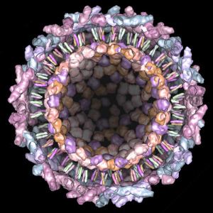
The Togaviridae family belong to group IV of the Baltimore classification of viruses. The genome is linear, non-segmented, single-stranded, positive sense RNA that is 10,000–12,000 nucleotides long. The 5'-terminus carries a methylated nucleotide cap and the 3'-terminus has a polyadenylated tail, therefore resembling cellular mRNA. The virus is enveloped and forms spherical particles (65–70 nm diameter), the capsid within is icosahedral, constructed of 240 monomers, having a triangulation number of 4.
Life Cycle
Entry into the host cell is achieved by attachment of the viral E glycoprotein to host receptors, which mediates clathrin-mediated endocytosis. The receptors for binding are unknown, however the tropism is varied and it is known that the glycoprotein petal-like spikes act as attachment proteins. After virus attachment and entry into the cell, gene expression and replication takes place within the cytoplasm.
Replication follows the positive stranded RNA virus replication model. Positive-stranded RNA-virus-transcription is the method of transcription. Translation takes place by viral initiation, and suppression of termination. The vector for Togaviridae is primarily the mosquito, where replication of the virus occurs. The Togaviridae family is classified into Old World and New World viruses based on geographical distribution, although it’s likely that a few transoceanic crossings have occurred. Human, mammals, marsupials, birds, and mosquitoes serve as the natural host. Transmission routes are zoonosis, bite, and respiratory.
Replication
The non-structural proteins are encoded at the 5’ end, formed during the first of two characteristic rounds of translation. These proteins are originally translated as a polyprotein, which consequently undergo self cleavage, forming four non-structural proteins responsible for gene expression and replication. The formation of a sub-genomic fragment, encoding the structural proteins and a negative sense fragment, a template for further synthesis of positive sense RNA are the characteristic second phase of translation. Assembly takes place at the cell surface, where the virus buds from the cell, acquiring the envelope. The replication cycle is very fast, taking around 4 hours.
History
Initially the Togavirus family included what are now called the Flaviviruses, within the Alphavirus genus. The Flaviviruses were formed into their own family when sufficient differences with the Alphaviruses were noted thanks to the development of sequencing.