
 | ||
Other names (Time-lapse) microcinematograph, (Time-lapse) video microscope, Time-lapse cinemicrograph Uses Observation of slow microscopic processes Inventor Jean Comandon and other contemporaries Related items | ||
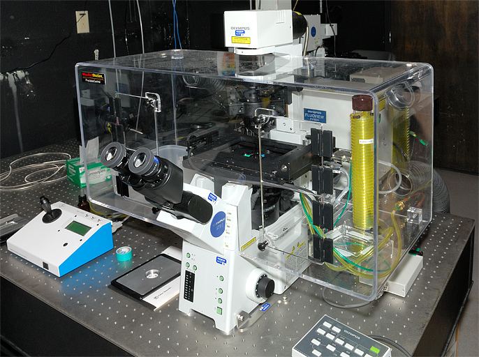
Time-lapse microscopy is time-lapse photography applied to microscopy. Microscope image sequences are recorded and then viewed at a greater speed to give an accelerated view of the microscopic process.
Contents
Before the introduction of the video tape recorder in the 1960s, time-lapse microscopy recordings were made on photographic film. During this period, time-lapse microscopy was referred to as microcinematography. With the increasing use of video recorders, the term time-lapse video microscopy was gradually adopted. Today, the term video is increasingly dropped, reflecting that a digital still camera is used to record the individual image frames, instead of a video recorder.
Applications
Time-lapse microscopy can be used to observe any microscopic object over time. However, its main use is within cell biology to observe artificially cultured cells. Depending on the cell culture, different microscopy techniques can be applied to enhance characteristics of the cells as most cells are transparent.
To enhance observations further, cells have therefore traditionally been stained before observation. Unfortunately, the staining process kills the cells. The development of less destructive staining methods and methods to observe unstained cells has led to that cell biologists increasingly observe living cells. This is known as live cell imaging.
Time-lapse microscopy is the method that extends live cell imaging from a single observation in time to the observation of cellular dynamics over long periods of time. Time-lapse microscopy is primarily used in research, but is clinically used in IVF clinics as studies has proven it to increase pregnancy rates, lower abortion rates and predict aneuploidy
Modern approaches are further extending time-lapse microscopy observations beyond making movies of cellular dynamics. Traditionally, cells have been observed in a microscope and measured in a cytometer. Increasingly this boundary is blurred as cytometric techniques are being integrated with imaging techniques for monitoring and measuring dynamic activities of cells and subcellular structures.
History
The Cheese Mites by Martin Duncan from 1903 is one of the earliest microcinematographic films. However, the early development of scientific microcinematography took place in Paris. The first reported time-lapse microscope was assembled in the late 1890s at the Marey Institute, founded by the pioneer of chronophotography, Étienne-Jules Marey. It was, however, Jean Comandon who made the first significant scientific contributions in around 1910.
Comandon was a trained microbiologist specializing in syphilis research. Inspired by Victor Henri's microcinematic work on Brownian motion, he used the newly invented ultramicroscope to study the movements of the syphilis bacteria. At the time, the ultramicroscope was the only microscope in which the thin spiral shaped bacteria was visible. Using an enormous cinema camera bolted to the fragile microscope, he demonstrated visually that the movement of the disease-causing bacteria is uniquely different from the non-disease-causing form. Comandon’s films proved instrumental in teaching doctors how to distinguish the two forms.
Comandon's extensive pioneering work inspired others to adopt microcinematography. Heniz Rosenberger builds a microcinematograph in the mid 1920s. In collerboration with Alexis Carrel, they used the device to further develop Carrel's cell culturing techniques. Similar work was conducted by Warren Lewis.
During the second World War II Carl Zeiss AG released the first phase contrast microscope on the market. With this new microscope, cellular details could for the first time be observed without using lethal stains. By setting up some of the first time-lapse experiments with chicken fibroblasts and a phase contrast microscope, Michael Abercrombie described the basis of our current understanding of cell migration in 1953.
With the broad introduction of the digital camera at the beginning of this century, time-lapse microscopy has been made dramatically more accessible and is currently experiencing an unrepresented raise in scientific publications.