
Entrez 7038 | Ensembl ENSG00000042832 | |
 | ||
Aliases TG, AITD3, TGN, thyroglobulin External IDs OMIM: 188450 MGI: 98733 HomoloGene: 2430 GeneCards: TG | ||
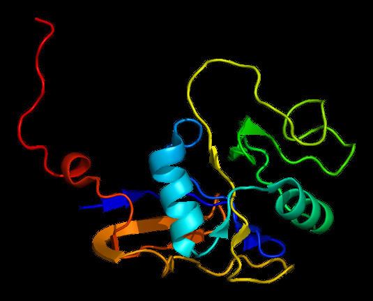
Thyroglobulin (Tg) is a 660 kDa, dimeric protein produced by the follicular cells of the thyroid and used entirely within the thyroid gland. Thyroglobulin protein accounts for approximately half of the protein content of the thyroid gland. Human TG is a homodimer of subunits each containing 2768 amino acids as synthesized (a short signal peptide may be removed from the N-terminus in the mature protein).
Contents
The protein is a precursor of the thyroid hormones; these are produced when thyroglobulin's tyrosine residues are combined with iodine and the protein is subsequently cleaved. Each thyroglobulin molecule contains approximately 100-120 tyrosine residues, but only a small number (20) of these are subject to iodination by thyroperoxidase in the follicular colloid. Therefore, each Tg molecule forms only approximately 10 thyroid hormone molecules.
Function
Tg is used by the thyroid gland to produce the thyroid hormones thyroxine (T4) and triiodothyronine (T3). The active form of triiodothyronine, 3, 5, 3' triiodothyronine, is produced both within the thyroid gland and in the periphery by 5'-deiodinase (which has been referred to as tetraiodothyronine 5' deiodinase). It is presumed that Tg and thyroid are also an important storage of iodine for all body needs, in particular, for many iodine-concentrating organs such as breast, stomach, salivary glands, thymus, choroid plexus and cerebrospinal fluid, etc. (see iodine in biology).
Tg is produced by the thyroid epithelial cells, called thyrocytes, which form spherical follicles. Tg is secreted and stored in the follicular lumen.
Via a reaction with the enzyme thyroperoxidase, iodine is covalently bound to tyrosine residues in thyroglobulin molecules, forming monoiodotyrosine (MIT) and diiodotyrosine (DIT).
Small globules of the follicular colloid (Tg) are endocytosed (hormone (TSH)-mediated) and proteases in lysosomes digest iodinated thyroglobulin, releasing T3 and T4 within the thyrocyte cytoplasm. The T3 and T4 are then transported across (TSH-mediated) the basolateral thyrocyte membrane, into the bloodstream, by an unknown mechanism, while the lysosome is recycled back to the follicular lumen.
Half-life and clinical elevation
Metabolism of thyroglobulin occurs in the liver and via thyroid gland recycling of the protein. Circulating thyroglobulin has a half-life of 65 hours. Following thyroidectomy, it may take many weeks before thyroglobulin levels become undetectable. After thyroglobulin levels become undetectable (following thyroidectomy), levels can be serially monitored.
A subsequent elevation of the thyroglobulin level is an indication of recurrence of papillary or follicular thyroid carcinoma. Hence, thyroglobulin levels in the blood are mainly used as a tumor marker for certain kinds of thyroid cancer (particularly papillary or follicular thyroid cancer). Thyroglobulin is not produced by medullary or anaplastic thyroid carcinoma.
Thyroglobulin antibodies
In the clinical laboratory, thyroglobulin testing can be complicated by the presence of anti-thyroglobulin antibodies (ATAs), alternatively referred to as TgAb. Anti-thyroglobulin antibodies are present in 1 in 10 normal individuals, and a greater percentage of patients with thyroid carcinoma. The presence of these antibodies can result in falsely low (or rarely falsely high) levels of reported thyroglobulin, a problem that can be somewhat circumvented by concomitant testing for the presence of ATAs. The ideal strategy for a clinician's interpretation and management of patient care in the event of confounding detection of ATAs is testing to follow serial quantitative measurements (rather than a single laboratory measurement).
ATAs are often found in patients with Hashimoto's thyroiditis or Graves' disease. Their presence is of limited use in the diagnosis of these diseases, since they may also be present in healthy euthyroid individuals. ATAs are also found in patients with Hashimoto's encephalopathy, a neuroendocrine disorder related to—but not caused by—Hashimoto's thyroiditis.
Interactions
Thyroglobulin has been shown to interact with Binding immunoglobulin protein.