
Artery Deep temporal arteries | Antagonist Platysma muscle | |
 | ||
Origin Temporal lines on the parietal bone of the skull and the superior temporal surface of the sphenoid bone. Insertion Coronoid process of the mandible. Nerve Deep temporal nerves, branch(es) of the anterior division of the mandibular nerve (V3) Actions Elevation and retraction of mandible | ||
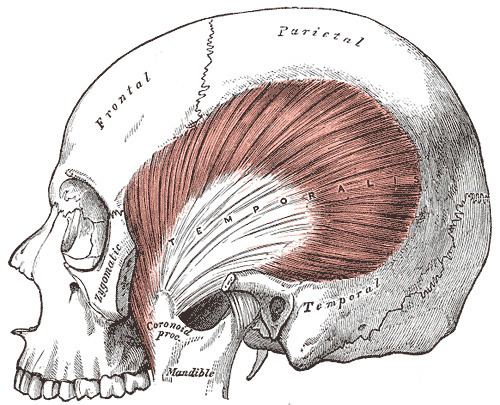
The temporal muscle, also known as the temporalis, is one of the muscles of mastication. It is a broad, fan-shaped muscle on each side of the head that fills the temporal fossa, superior to the zygomatic arch so it covers much of the temporal bone.
Contents
Structure
In humans, it arises from the temporal fossa and the deep part of temporal fascia. It passes medial to the zygomatic arch and forms a tendon which inserts onto the coronoid process of the mandible, with its insertion extending into the retromolar fossa posterior to the most distal mandibular molar. In other mammals, the muscle usually spans the dorsal part of the skull all the way up to the medial line. There, it may be attached to a sagittal crest, as can be seen in early hominins like Paranthropus aethiopicus.
The temporal muscle is covered by the temporal fascia, also known as the temporal aponeurosis. This fascia is commonly used in tympanoplasty, or surgical reconstruction of the eardrum.
The muscle is accessible on the temples, and can be seen and felt contracting while the jaw is clenching and unclenching.
Development
The temporalis is derived from the first pharyngeal arch in development.
Innervation
As with the other muscles of mastication, control of the temporal muscle comes from the third (mandibular) branch of the trigeminal nerve. Specifically, the muscle is innervated by the deep temporal nerves.
Blood supply
The muscle receives its blood supply from the deep temporal arteries which anastomose with the middle temporal artery.
Function
The temporal muscle is the most powerful muscle of the temporomandibular joint. The temporal muscle can be divided into two functional parts; anterior and posterior. The anterior portion runs vertically and its contraction results in elevation of the mandible (closing the mouth). The posterior portion has fibers which run horizontally and contraction of this portion results in retrusion of the mandible.
When lower dentures are fitted, they should not extend into the retromolar fossa to prevent trauma of the mucosa due to the contraction of the temporalis muscle.
Pathology
The temporalis is likely to be involved in jaw pain and headaches. Bruxism, the habitual grinding of teeth typically while sleeping, and clenching of the jaw while stressed can lead to overwork of the temporalis and results in pain. A myotendinous rupture of the temporalis can occur during a seizure due to extreme clenching of the jaw. During a seizure the contralateral temporalis muscle can enter spastic paralysis, this clenching in extreme cases can lead to a rupture specifically on the myotendinous insertion at the coronoid process of the mandible.