
 | ||
Talairach coordinates, also known as Talairach space, is a 3-dimensional coordinate system (known as an 'atlas') of the human brain, which is used to map the location of brain structures independent from individual differences in the size and overall shape of the brain. It is still common to use Talairach coordinates in functional brain imaging studies and to target transcranial stimulation of brain regions. However, alternative methods such as the MNI Coordinate System originated at the Montreal Neurological Institute and Hospital have largely replaced Talairach for stereotaxy and other procedures.
Contents
History
The coordinate system was first created by neurosurgeons Jean Talairach and Gabor Szikla in their work on the Talairach Atlas in 1967, creating a standardized grid for neurosurgery. The grid was based on the idea that distances to lesions in the brain are proportional to overall brain size (i.e., the distance between two structures is larger in a larger brain). In 1988 a second edition of the Talairach Atlas came out that was coauthored by Tournoux, and it is sometimes known as the Talairach-Tournoux system. This atlas was based on single post-mortem dissection of a human brain.
The Talairach Atlas uses Brodmann areas as the labels for brain regions.
Description
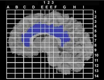
The Talairach coordinate system is defined by making two anchors, the anterior commissure and posterior commissure, lie on a straight horizontal line. Since these two points lie on the midsagittal plane, the coordinate system is completely defined by requiring this plane to be vertical. Distances in Talairach coordinates are measured from the anterior commissure as the origin (as defined in the 1998 edition). The y-axis points posterior and anterior to the commissures, the left and right is the x-axis, and the z-axis is in the ventral-dorsal (down and up) directions. Once the brain is reoriented to these axes, the researchers must also outline the six cortical outlines of the brain: anterior, posterior, left, right, inferior, and superior. In the 1967 atlas the left is with positive coordinates while in the 1988 atlas the left has negative coordinates.
By defining standard anatomical landmarks that could be identified on different subjects (the anterior and posterior commissures), it became easier to spatially warp an individual brain image obtained through Magnetic Resonance Imaging (MRI), positron emission tomography (PET) and other imaging methods to this standard Talairach space. One can then make inferences about tissue identity at a specific location by referring to the atlas.
Conversion to other coordinate systems
Another common atlas for the human brain is the Montreal Neurological Institute and Hospital (MNI) coordinate system, which is the template used for SPM and the International Consortium for Brain Mapping. Most neuroimaging software packages are able to convert from Talairach to MNI coordinates.