
Symbol TCR_zetazeta InterPro IPR021663 OPM protein 2hac | Pfam PF11628 OPM superfamily 261 Pfam structures | |
 | ||
The T-cell receptor, or TCR, is a molecule found on the surface of T cells, or T lymphocytes, that is responsible for recognizing fragments of antigen as peptides bound to major histocompatibility complex (MHC) molecules. The binding between TCR and antigen peptides is of relatively low affinity and is degenerate: that is, many TCRs recognize the same antigen peptide and many antigen peptides are recognized by the same TCR.
Contents
- Structural characteristics of the TCR
- Generation of the TCR diversity
- The TCR complex
- TCR co receptors
- Associated molecules of the TCR complex involved in T cell activation
- References
The TCR is composed of two different protein chains (that is, it is a heterodimer). In humans, in 95% of T cells the TCR consists of an alpha (α) chain and a beta (β) chain (encoded by TRA and TRB, respectively), whereas in 5% of T cells the TCR consists of gamma and delta (γ/δ) chains (encoded by TRG and TRD, respectively). This ratio changes during ontogeny and in diseased states (such as leukemia). It also differs between species. Orthologues of the 4 loci have been mapped in various species. Each locus can produce a variety of polypeptides with constant and variable regions.
When the TCR engages with antigenic peptide and MHC (peptide/MHC), the T lymphocyte is activated through signal transduction, that is, a series of biochemical events mediated by associated enzymes, co-receptors, specialized adaptor molecules, and activated or released transcription factors.
Structural characteristics of the TCR
The TCR is a disulfide-linked membrane-anchored heterodimeric protein normally consisting of the highly variable alpha (α) and beta (β) chains expressed as part of a complex with the invariant CD3 chain molecules. T cells expressing this receptor are referred to as α:β (or αβ) T cells, though a minority of T cells express an alternate receptor, formed by variable gamma (γ) and delta (δ) chains, referred as γδ T cells.
Each chain is composed of two extracellular domains: Variable (V) region and a Constant (C) region, both of Immunoglobulin superfamily (IgSF) domain forming antiparallel β-sheets. The Constant region is proximal to the cell membrane, followed by a transmembrane region and a short cytoplasmic tail, while the Variable region binds to the peptide/MHC complex.
The variable domain of both the TCR α-chain and β-chain each have three hypervariable or complementarity determining regions (CDRs). There is also an additional area of hypervariability on the β-chain (HV4) that does not normally contact antigen and, therefore, is not considered a CDR.
The residues in these variable domains are located in two regions of the TCR, at the interface of the α- and β-chains and in the β-chain framework region that is thought to be in proximity to the CD3 signal-transduction complex. CDR3 is the main CDR responsible for recognizing processed antigen, although CDR1 of the alpha chain has also been shown to interact with the N-terminal part of the antigenic peptide, whereas CDR1 of the β-chain interacts with the C-terminal part of the peptide.
CDR2 is thought to recognize the MHC. CDR4 of the β-chain is not thought to participate in antigen recognition, but has been shown to interact with superantigens.
The constant domain of the TCR consists of short connecting sequences in which a cysteine residue forms disulfide bonds, which form a link between the two chains.
The TCR is a member of the immunoglobulin superfamily, a large group of proteins involved in binding, recognition, and adhesion; the family is named after antibodies (also called immunoglobulins). The TCR is similar to a half-antibody consisting of a single heavy and single light chain, except the heavy chain is without its crystallisable fraction (Fc). The two subunits of TCR are twisted together. Whereas the antibody uses its Fc region to bind to Fc Receptors on leukocytes, TCR is already docked onto the cell membrane. However, it is not able to mediate signal transduction itself due to its short cytoplasmic tail, so TCR still requires CD3 and zeta to carry out the signal transduction in its place, just as antibodies require binding to FcRs to initiate signal transduction. In this way the MHC-TCR-CD3 interaction for T cells is functionally similar to the antigen(Ag)-immunoglobulin(Ig)-FcR interaction for myeloid leukocytes, and Ag-Ig-CD79 interaction for B cells.
Generation of the TCR diversity
Processes for the generation of TCR diversity are similar to those described for antibodies and B cell antigen receptors. It is based mainly on genetic recombination of the DNA encoded segments in individual somatic T cells – either somatic V(D)J recombination using RAG1 and RAG2 recombinases. Unlike immunoglobulins, TCRs do not undergo somatic hypermutation and do not express Activation-Induced (Cytidine) Deaminase (AID). The recombination process that creates diversity in BCR (antibodies) and TCR is unique to lymphocytes (T and B cells) during the early stages of their development in primary lymphoid organs (thymus for T cells, bone marrow for B cells).
Each recombined TCR possess unique antigen specificity, determined by the structure of the antigen-binding site formed by the α and β chains in case of αβ T cells or γ and δ chains on case of γδ T cells.
The intersection of these specific regions (V and J for the alpha or gamma chain; V, D, and J for the beta or delta chain) corresponds to the CDR3 region that is important for peptide/MHC recognition (see above).
It is the unique combination of the segments at this region, along with palindromic and random nucleotide additions (respectively termed "P-" and "N-"), which accounts for the even greater diversity of T-cell receptor specificity for processed antigenic peptides.
Later during development, individual CDR loops of TCR can be re-edited in the periphery outside thymus by reactivation of recombinases using a process termed TCR revision (editing) and change its antigenic specificity.
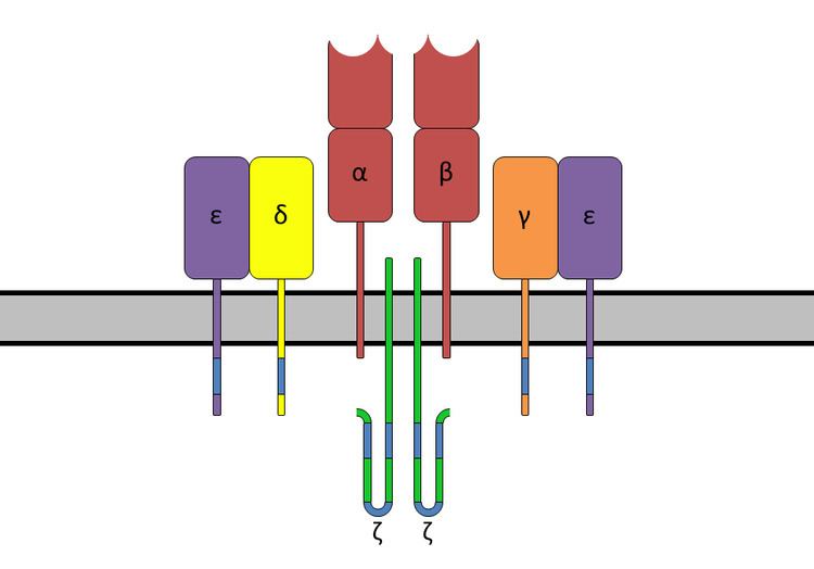
The TCR complex
The TCR receptor complex is an octomeric complex of variable TCR receptor α and β chains with three dimeric signaling modules CD3δ/ε, CD3γ/ε and CD247 ζ/ζ or ζ/η. Ionizable residues in the transmembrane domain of each subunit form a polar network of interactions that hold the complex together. Since the cytoplasmic tail of the TCR is extremely short, making it unlikely to participate in signaling, these signaling molecules are vital in propagating the signal from the triggered TCR into the cell.
Each T cell expresses clonal TCRs which recognize specific peptide/MHC complex during physical contact between T cell and antigen-presenting cell-APC (MHC class II) or any other cell type (MHC class I) High on-rate and off-rate is characteristic for TCR and peptide/MHC interaction at physiological temperature. TCRs have very high degree of antigen specificity, despite of fact that the affinity to the peptide/MHC ligand is in the micromolar range. This weak binding (
TCR co-receptors
The signal from the T-cell complex is enhanced by simultaneous binding of the MHC molecules by a specific co-receptor.
Extracellularly, the TCR co-receptor defines the specificity of the TCR to MHC class I or II molecule, and increases binding affinity of TCR to MHC to prolong the cell-cell interaction between the antigen-presenting cell and the T cell.
Intracellularly, the TCR co-receptor recruits essential molecules (e.g., LCK) involved in the signaling of the activated T lymphocyte to facilitate the CD3 signal transduction mechanism.
Associated molecules of the TCR complex involved in T-cell activation
The essential function of the TCR complex is to identify specific bound antigen and elicit a distinct and critical response. The signal transduction mechanism by which a T cell elicits this response upon contact with its unique antigen is termed T-cell activation (just as phototransduction is the term given to the signal transduction event by which photoreceptors elicits vision upon exposure to photons). There are myriad molecules involved in the complex biochemical process (called trans-membrane signaling) by which T-cell activation occurs.
The most common mechanism for activation and regulation of molecules beneath the lipid bilayer is via reversible tyrosine phosphorylation by protein kinase/phosphatase. T cells utilise the Src family kinases in transmembrane signalling largely to phosphorylate tyrosines that are part of immunoreceptor tyrosine-based activation motifs (ITAM) in intracellular parts of CD3 and ζ chains.
Early signaling steps implicate the following kinases and phosphatases after TCR triggering:
When a T-cell receptor is activated by contact with a peptide:MHC complex, CD45 dephosphorylates inhibitory tyrosine of membrane-localized Src family kinases Fyn and Lck, previously recruited and activated by CD4 or CD8 coreceptors. Activated Fyn and Lck phosphorylates ITAMs on the CD3 and ζ chains. This allows cytoplasmic kinases of the Syk family (ZAP-70) to bind to the ITAM and activated ZAP-70 phosphorylates tyrosines on the adaptor protein LAT, which then attracts PLC-γ. Other downstream pathways are triggered as well (MAPK, NF-κB, NFAT) which results in gene transcription in the nucleus.