
TA A14.1.09.006 | ||
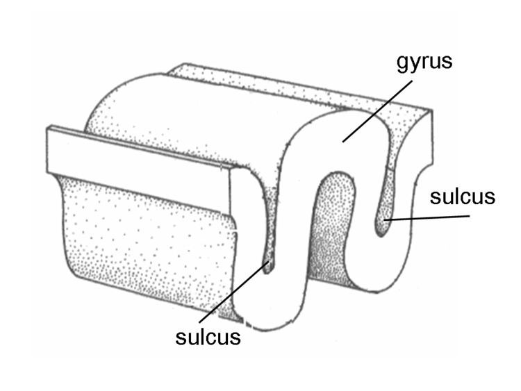 | ||
In neuroanatomy, a sulcus (Latin: "furrow", pl. sulci) is a depression or groove in the cerebral cortex. It surrounds a gyrus (pl. gyri), creating the characteristic folded appearance of the brain in humans and other mammals.
Contents
Structure
Sulci are one of three parts of the cerebral cortex, the others being the gyri and the fissures. The three different parts create a larger surface area for the human brain and other mammalian brains. When looking at the human brain, two-thirds of the surface are hidden in the grooves. The sulci and fissures are both grooves in the cortex but they are differentiated by size. A sulcus is a shallower groove that surrounds a gyrus. A fissure is a large furrow that divides the brain into lobes, and also into the two hemispheres as the medial longitudinal fissure does. However this distinction is not always clear. For example, the lateral sulcus is also known as the lateral fissure or the Sylvian fissure, and the central sulcus is also known as the central fissure or the Rolandic fissure.
Importance of expanded surface area
As the surface area of the brain increases more functions are made possible. A smooth-surfaced brain is only able to grow to a certain extent. A depression, sulcus, in the surface area allows for continued growth. This in turn allows for the functions of the brain to continue growing.
Variation
The sulcal pattern varies between human individuals, and the most elaborate overview on this variation is probably an atlas by Ono, Kubick and Abernathey: Atlas of the Cerebral Sulci. Some of the more prominent sulci are, however, seen across individuals - and even species - making a common nomenclature across individuals and species possible.
Development
In humans, cerebral convolutions appear at about 5 months and take at least into the first year after birth to fully develop. Development varies greatly between individuals. The potential influences of genetic, epigenetic and environmental factors are not fully understood. It has been found that the width of cortical sulci not only increases with age, but also with cognitive decline in the elderly.
Notable sulci
Other animals
The variation in the amount of fissures in the brain (gyrification) between species is related to the size of the animal and the size of the brain. Mammals that have smooth-surfaced or nonconvoluted brains are called lissencephalics and those that have folded or convoluted brains gyrencephalics. The division between the two groups occurs when cortical surface area is about 10 cm2 and the brain has a volume of 3–4 cm3. Large rodents such as beavers (40 pounds (18 kg)) and capybaras (150 pounds (68 kg)) are gyrencephalic and smaller rodents such as rats and mice lissencephalic.
Macaque
A macaque has a more simple sulcal pattern. In a monograph Bonin and Bailey list the following as the primary sulci: