
Latin sinus sphenoidalis Dorlands
/Elsevier s_12/12739248 FMA 54683 | MeSH A04.531.621.827 TA A06.1.03.003 | |
 | ||
Nerve posterior ethmoidal nerves, and orbital branches of the pterygopalatine ganglion | ||
Each of the paired sphenoidal sinuses (components of the paranasal sinuses) is contained within the body of the sphenoid. They vary in size and shape and owing to the lateral displacement of the intervening septum they are rarely symmetrical. They cannot be palpated during an extraoral examination.
Contents
The following are their average measurements: vertical height, 2.2 cm.; transverse breadth, 2 cm.; antero-posterior depth, 2.2 cm.
Relations
When exceptionally large they may extend into the roots of the pterygoid processes or great wings, and may invade the basilar part of the occipital bone.
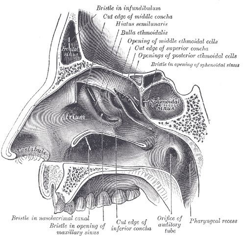
Each sinus opens into the roof of the nasal cavity via apertures on the posterior wall of the sphenoethmoidal recess directly above the choana. The apertures are located high on the anterior walls of the sinuses themselves.
Development
They are present as very small cavities at birth, and slowly develop with the growth of the skull. Just after puberty the sinuses finish development.
Innervation
The mucous membrane receives sensory innervation by the posterior ethmoidal nerves (branch of the ophthalmic nerve), and postganglionic parasympathetic fibers of the facial nerve that synapsed at the pterygopalatine ganglion which control secretion of mucus.
Complications
If the tumor spreads laterally, the cavernous sinus and all its constituent nerves could be in danger.
Use in neurosurgery
Because only thin shelves of bone separate the sphenoidal sinuses from the nasal cavities below and hypophyseal fossa above, the pituitary gland can be surgically approached through the roof of the nasal cavities by first passing through the anterioinferior aspect of the sphenoid bone and into the sinuses, followed by entry through the top of the sphenoid bone into the hypophyseal fossa.