
Latin nuclei septales Dorlands
/Elsevier n_11/12583395 TA A14.1.09.266 | NeuroNames hier-241 BAMS SEPTAL-NUCLEI FMA 61845 | |
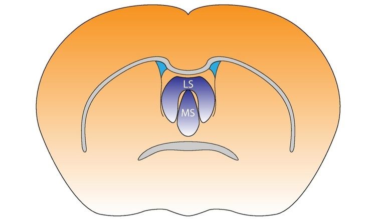 | ||
The septal nuclei (medial olfactory area) are a set of structures that lie below the rostrum of the corpus callosum, anterior to the lamina terminalis (the layer of gray matter in the brain connecting the optic chiasma and the anterior commissure where the latter becomes continuous with the rostral lamina). The septal nuclei are composed of medium-size neurons which are classified into medial, lateral, and posterior groups. The septal nuclei receive reciprocal connections from the olfactory bulb, hippocampus, amygdala, hypothalamus, midbrain, habenula, cingulate gyrus, and thalamus. The septal area (medial olfactory area) has no relation to the sense of smell, but it is considered a pleasure zone in animals. The septal nuclei play a role in reward and reinforcement along with the nucleus accumbens. In the 1950s, Olds & Milner showed that rats with electrodes implanted in this area will self-stimulate repeatedly (i.e. press a bar to receive electrical current that will stimulate the neurons). Experiments on the septal area of man have taken place since the 1960s.
Contents
Connections with other structures
The dorsal septum projects to the lateral preoptic area, lateral hypothalamus, periventricular hypothalamus and midline thalamus.
Fibers from the ventral half of the septum project topographically to the hippocampal formation, thalamus, hypothalamus and midbrain. Specifically, neurons located along the midline in the vertical limb of the diagonal band of Broca project through the dorsal fornix to all CA fields of the dorsal hippocampus and adjacent subicular cortex. Other fibers from this region project through the stria medullaris to the medial and lateral habenular nuclei, the paratenial and anteromedial nucleus of the thalamus, and through the medial forebrain bundle to the pars posterior of the medial mammillary nucleus.
Cells located in the intermediolateral septum also project through the lateral part of the fimbria to all CA fields of the ventral hippocampus and adjacent subicular and entorhinal cortices. These cells also send fibers through the stria medullaris to the lateral habenular nucleus and mediodorsal thalamic nucleus. Other axons arising from these cells descend through the medial forebrain bundle to terminate in a region dorsal to the interpeduncular nucleus.
The lateral septum is a relay center for connections from the CA3 of the hippocampus to the ventral tegmental area. These connections help link reward signals with the context in which they occur.
Fibers from the most lateral part of the ventral septum (i.e., bed nucleus of the anterior commissure) project through the stria terminalis to the ventral subiculum. In addition, cells located in the horizontal limb of the diagonal band project massively to the pars posterior of the medial mammillary nucleus, the ventral tegmental area, and amygdala.
Lateral Septum and Social Behavior
GABA and Glutamate which regulates Lateral Septum (LS) activity has been found to be increased during social play in rats. No sex differences were found in extracellular GABA concentrations during social playing, however; Glutamate plays a major role in female social playing. When Glutamate receptors are blocked in the LS pharmacologically, there is a significant decrease in female social playing, while males had no decrease in playing. This suggests that GABA neurotransmission in the LS is involved in social play behavior regulation in both sexes, while glutamate neurotransmission in the LS is involved in sex-specific regulation of social play in rats.