
Group Group V ((−)ssRNA) Family Paramyxoviridae Scientific name Sendai virus Rank Species | Order Mononegavirales Genus Respirovirus Higher classification Respirovirus | |
 | ||
Similar Measles virus, Double‑stranded RNA viruses, Respirovirus, Ectromelia virus, Rubulavirus | ||
How does sendai virus reprogram cells
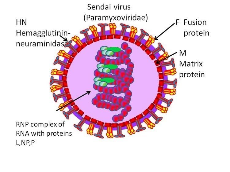
Sendai virus (SeV), previously also known as murine parainfluenza virus type 1 or hemagglutinating virus of Japan (HVJ), is a negative sense, single-stranded RNA virus of the family Paramyxoviridae, a group of viruses featuring, notably, the genera Morbillivirus and Rubulavirus. SeV is a member of genus Respirovirus, members of which primarily infect mammals.
Contents
- How does sendai virus reprogram cells
- Oncolytic sendai virus
- Diagnosis and Prophylaxis
- Symptoms
- Sendai Fusion
- References
SeV is responsible for a highly transmissible respiratory tract infection in mice, hamsters, guinea pigs, rats, and occasionally pigs, and marmosets with infection passing through both air and direct contact routes. The virus can be detected in mouse colonies worldwide, generally in suckling to young adult mice. Epizootic infections of mice are usually associated with a high mortality rate, while enzootic disease patterns suggest that the virus is latent and can be cleared over the course of a year. Sublethal exposure to SeV can promote long-lasting immunity to further lethal doses of SeV.
A novel and well-recognized use for SeV is the fusion of eukaryotic cells, especially to produce hybridoma cells capable of manufacturing monoclonal antibodies in large quantities.
Oncolytic sendai virus
Diagnosis and Prophylaxis
SeV induces lesions within the respiratory tract, usually associated with bacterial inflammation of the trachea and lung (tracheitis and bronchopneumonia, respectively). However, the lesions are limited, and aren't indicative solely of SeV infection. Detection, therefore, makes use of SeV-specific antigens in several serological methods, including ELISA, immunofluorescence, and hemagglutination assays, with particular emphasis on use of the ELISA for its high sensitivity (unlike the hemagglutination assay) and its fairly early detection (unlike the immunofluorescence assay).
In a natural setting, the respiratory infection of Sendai virus in mice is acute. From the extrapolation of the infection of laboratory mice, the presence of the virus may first be detected in the lungs 48 to 72 hours following exposure. As the virus replicates in the respiratory tract of an infected mouse, the concentration of the virus grows most quickly during the third day of infection. After that, the growth of the virus is slower but consistent. Typically, the peak concentration of the virus is on the sixth or seventh day, and rapid decline follows that by the ninth day. A fairly vigorous immune response mounted against the virus is the cause of this decline. The longest period of detected presence of the virus in a mouse lung is fourteen days after infection.
Eaton et al. advises that, when controlling an outbreak of SeV, disinfecting the laboratory environment and vaccinating the breeders, as well as eliminating infected animals and screening incoming animals, should clear the problem very quickly. Imported animals should be vaccinated with SeV and placed in quarantine, while, in the laboratory environment, breeding programs should be discontinued, and the non-breeding adults isolated for two months.
Symptoms
Sendai Fusion
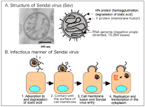
One recognized feature of the Sendai virus, shared with members of its genus, is the ability to induce syncytia formation in vitro in eukaryotic colonies. The mechanism for this process is fairly well understood and is very similar to the fusion process employed by the virion to facilitate cellular entry. The activities of the receptor binding hemagglutinin-neuraminidase protein is solely responsible for inducing close interaction between the virus envelope and the cellular membrane.
However, it is the F protein (one of many membrane fusion proteins) that, when triggered by local dehydration and a conformation change in the bound HN protein, actively inserts into the cellular membrane, which causes the envelope and the membrane to merge, followed shortly by virion entry. When the HN and F protein are manufactured by the cell and expressed on the surface, the same process may occur between adjacent cells, causing extensive membrane fusion and resulting in the formation of a syncytium.
This behavior of SeV was utilized by Köhler and Milstein, who published an article in 1975 outlining a revolutionary method of manufacturing monoclonal antibodies. In need of a reliable method to produce large quantities of a specific antibody, the two merged a monoclonal B cell, exposed to a chosen antigen, and a myeloma tumor cell to produce hybridomas, capable of being grown indefinitely and of producing significant amounts of an antibody specifically targeting the chosen antigen. Though more efficient methods of creating such hybrids have since been found, Köhler and Milstein first used Sendai virus to create their revolutionary cells.