
Precursor Wolffian duct MeSH A05.360.444.713 | Latin Vesiculae seminales FMA 19386 | |
 | ||
Artery Inferior vesical artery, middle rectal artery Lymph External iliac lymph nodes, internal iliac lymph nodes | ||
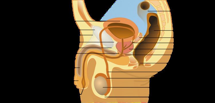
The seminal vesicles (Latin: glandulae vesiculosae), vesicular glands, or seminal glands, are a pair of simple tubular glands posteroinferior to the urinary bladder of some male mammals. Seminal vesicles are located within the pelvis. They secrete fluid that partly composes the semen.
Contents
Structure
The seminal vesicles are a pair of glands that are positioned below the urinary bladder and lateral to the vas deferens. Each vesicle consists of a single tube folded and coiled on itself, with occasional diverticula in its wall.
The excretory duct of each seminal gland unites with the corresponding vas deferens to form the two ejaculatory ducts, which immediately pass through the substance of the prostate gland before opening separately into the verumontanum of the prostatic urethra.
Each seminal vesicle spans approximately 5 cm, though its full unfolded length is approximately 10 cm, but it is curled up inside the gland's structure.
Development
Each vesicle forms as an outpocketing of the wall of the ampulla of one vas deferens. The seminal vesicles develop as one of three structures of the male reproductive system that develops at the junction between the urethra and vas deferens. The vas deferens is derived from the mesonephric duct, a structure that develops from mesoderm.
Histology
Under microscopy, the seminal vesicles can be seen to have a mucosa, consisting of a lining of interspersed columnar cells and a lamina propria; and a thick muscular wall. The lumen of the glands is highly irregular and stores secretions from the glands of the vesicles. In detail:
The height of these columnar cells, and therefore activity, is dependent upon testosterone levels in the blood.
Spermatozoa may occasionally be found within the lumen of the glands, even though the vesicles are blind-ended in nature. This is thought to be because of slight reflux due to muscular contractions of the urethra during ejaculation.
Function
The seminal vesicles secrete a significant proportion of the fluid that ultimately becomes semen. Lipofuscin granules from dead epithelial cells give the secretion its yellowish color. About 70-85% of the seminal fluid in humans originates from the seminal vesicles, but is not expelled in the first ejaculate fractions which are dominated by spermatozoa and zinc-rich prostatic fluid. The excretory duct of each seminal gland opens into the corresponding vas deferens as it enters the prostate gland. Seminal vesicle fluid is alkaline, resulting in human semen having a mildly alkaline pH. The alkalinity of semen helps neutralize the acidity of the vaginal tract, prolonging the lifespan of sperm. Acidic ejaculate (pH <7.2) may be associated with ejaculatory duct obstruction. The vesicle produces a substance that causes the semen to become sticky and jelly-like after ejaculation.
The thick secretions from the seminal vesicles contain proteins, enzymes, fructose, mucus, vitamin C, flavins, phosphorylcholine and prostaglandins. The high fructose concentrations provide nutrient energy for the spermatozoa when stored in semen in the laboratory.
In vitro studies have shown that sperm expelled together with seminal vesicular fluid show poor motility and survival, and the sperm chromatin is less protected. Therefore, the exact physiological importance of seminal vesicular fluid is not clear.
The development and maintenance of the seminal vesicles, as well as their secretion and size/weight, are highly dependent on androgens. The seminal vesicles contain 5α-reductase, which metabolizes testosterone into its much more potent metabolite, dihydrotestosterone (DHT). The seminal vesicles have also been found to contain luteinizing hormone receptors, and hence may also be regulated by the ligand of this receptor, luteinizing hormone.
Clinical significance
Physical examination of the seminal vesicles is difficult. Laboratory examination of seminal vesicle fluid requires a semen sample, e.g. for semen culture or semen analysis. Fructose levels provide a measure of seminal vesicle function and, if absent, bilateral agenesis or obstruction is suspected.
Disorders of the seminal vesicles include seminal vesiculitis, acquired cysts, abscess, congenital anomalies (such as agenesis, hypoplasia and cysts), amyloidosis, tuberculosis, schistosomiasis, hydatid cyst, calculi (stones) and tumours.
Primary adenocarcinoma of the seminal vesicles, although rare, constitutes the most common neoplasm of the seminal vesicles; even rarer neoplasms include sarcoma, squamous cell carcinoma, yolk sac tumor, neuroendocrine carcinoma, paraganglioma, epithelial stromal tumors and lymphoma.
Inflammation
Seminal vesiculitis (also known as spermatocystitis) is an inflammation of the seminal vesicles, most often caused by bacterial infection. Symptoms of seminal vesiculitis can include vague back or lower abdominal pain; penile, scrotal, or perineal pain; painful ejaculation; hematospermia; irritative and obstructive voiding symptoms; and impotence.
It is usually treated by administration of antibiotics. In intractable cases, in case of patient discomfort, transurethral seminal vesiculoscopy may be considered.