
ICD-10 S82.1 | AO 41-B1 | |
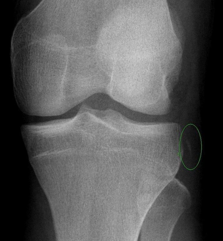 | ||
The Segond fracture is a type of avulsion fracture (soft tissue structures tearing off bits of their bony attachment) of the lateral tibial condyle of the knee, immediately beyond the surface which articulates with the femur.
Contents
History and incidence
Originally described by Dr. Paul Segond in 1879 after a series of cadaveric experiments, the Segond fracture occurs in association with tears of the anterior cruciate ligament (ACL) (75–100%) and injury to the medial meniscus (66–75%), lateral capsular ligament (now known as the Anterolateral ligament, or ALL), as well as injury to the structures behind the knee.
A rare, mirror image of the Segond fracture has also been described. The so-called "reverse Segond fracture" can occur after an avulsion fracture of the tibial component of the medial collateral ligament (MCL) in association with posterior cruciate ligament (PCL) and medial meniscal tears.
Segond fracture is typically the result of abnormal varus, or "bowing", stress to the knee, combined with internal rotation of the tibia. Reverse Segond fracture, as its name suggests, is caused by abnormal valgus, or "knock-knee", stress and external rotation.
Originally thought to be a result of avulsion of the medial third of the lateral collateral ligament, the Segond fracture has been shown by more recent research to relate also to the insertion of the iliotibial tract (ITT) and the anterior oblique band (AOB), a ligamentous attachment of the fibular collateral ligament (FCL), to the midportion of the lateral tibia and to be associated with avulsion by the anterolateral ligament (ALL). (Roberts CC, Towers JD, Spangehl MJ et-al. Advanced MR imaging of the cruciate ligaments. Radiol. Clin. North Am. 2007;45 (6): 1003-16, vi-vii.)
Clinical significance
Because of the high rate of associated ligamentous and meniscal injury, the presence of a Segond or reverse Segond fracture requires that these other pathologies must be specifically ruled out. Increasingly, reconstruction of the ACL is combined with reconstruction of the ALL when this associated pathology is present. It is often associated with an increased 'pivot shift' on physical exam.
Imaging findings
Segond and reverse Segond fractures are characterized by a small avulsion, or "chip", fragment of characteristic size that is best seen on plain radiography in the anterior-posterior plane. The chip of bone may be very difficult to see on the plain x-ray exam, and may be better seen on computed tomography. MRI may be useful for visualization of the associated bone marrow edema of the underlying tibial plateau on fat- saturated T2W and STIR images, as well as the associated findings of ligamentous and/or meniscal injury.