
Days 28 | Code TE E5.5.3.0.0.0.3 | |
 | ||
Precursor Ventral part of the Foregut Latin Gemma respiratoria,
gemma pulmonalis | ||
The respiratory bud is an embryological structure of endodermal origin that develops into organs of the respiratory system, such as the larynx, trachea and lungs.
Contents
Early stage
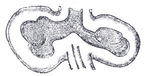
In the fourth week of development, the respiratory bud or diverticulum, starts to grow from the ventral (front) side of the foregut into the mesoderm that surrounds it, forming the lung bud. Around the 28th day, the lung bud splits into two bronchial buds that develop on the sides of the distal part of the respiratory bud and begin to grow laterally, expanding into the primitive pleural cavity.
Molecular signaling
The molecular signaling involved in the specification of the respiratory bud starts with the expression of the Nbx2-1 gene, which determines the respiratory field – the area where the respiratory bud will begin to grow from. The signaling that makes the growth of the respiratory bud possible is complex and involves a number of interactions between the mesoderm and the respiratory bud epithelium, in which members of the Fgf and Fgfr family of genes express.
Separation of trachea and esophagus
At first, the posterior part of the trachea is open to the esophagus, but as the bud elongates two longitudinal mesodermal ridges begin to form and grow until they join, forming a wall between the two organs. An incomplete separation of the organs leads to a congenital abnormality known as a tracheoesophageal fistula.
Larynx development
The epithelium of the larynx is of endodermal origin, but the laryngeal cartilages, unlike the rest of the respiratory bud connective tissue, come from the mesenchyme of the fourth and sixth pharyngeal arches. The fourth pharyngeal arch, adjacent to what will be the root of the tongue, will become the epiglottis. The sixth pharyngeal arch, located around the laryngeal orifice, will become the thyroid, cricoid and arytenoid cartilages. These structures are formed in a process in which the lining cells of the primitive larynx proliferate and occlude it. Later, it recanalizes leaving two membrane-like structures: the vocal folds and the vestibular folds. In between, an enlarged space, the ventricle, remains. Failure in this process leads to a serious but rare condition called congenital atresia of the larynx.
Later development
After the lung buds have formed, they begin to grow and branch forming a primitive version of the bronchial tree, determining how the lobes of the lung will be arranged in the mature organ. The first stage of alveolar development, spanning between the fifth and the 16th week of development, is called the pseudoglandular stage. It is so called because of the histological appearance of the primitive alveoli, which resemble glandular tissue. After the pseudoglandular stage, the lung enters the canalicular and saccular phases. During these stages, the terminal tubes narrow and give rise to small saccules, which become increasingly associated with capillaries as to make gas exchange possible. The alveolar epithelium begins to differentiate into two distinct types of cells: type I pneumocytes and type II pneumocytes, as well as the respiratory epithelium of the trachea and bronchial tree.