
 | ||
Reference Point Indentation (RPI) refers to a specialized form of indentation testing. RPI utilizes a unique method of measurement by establishing a relative reference point at the location of measurement. This unique capability makes it possible to measure materials that are in motion, oddly shaped, visco-elastic, or that may be coated or covered by another, softer material.
Contents
Unlike traditional indentation testing, RPI testing uses the location of measurement as the relative displacement reference position. Indentation itself is perhaps the most commonly applied means of testing the mechanical properties of materials. The technique has its origins in the Mohs scale of mineral hardness, in which materials are ranked according to what they can scratch and are, in turn, scratched by. The characterization of solids in this way takes place on an essentially discrete scale, so much effort has been expended in order to develop techniques for evaluating material hardness over a continuous range. Hence, the adoption of the Meyer, Knoop, Brinell, Rockwell, and Vickers hardness tests. More recently (ca. 1975), nanoindentation techniques have been established as the primary tool for investigating the hardness of small volumes of material. However, even more recently (ca. 2006), interest in measuring functional roles of biomaterials drove the development of the Reference Point Indentation technique.
New research in field such as biomaterials has led scientists to begin considering materials as complex systems that behave differently than the constituent parts. For example, materials like bone are hierarchical and made of many components including calcium, collagen, water, and non-collagenous proteins. Each of these components has unique material properties. When combined to form bone, the function of the tissue is different than any one constituent. Understanding this mechanical system is becoming a new field of research called Materiomics. RPI specifically aims to aid materiomics researchers understand the functional capabilities of these types of materials at a relevant length-scale.
Background
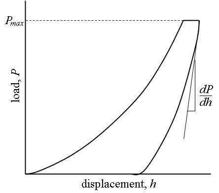
In a traditional indentation test (macro or micro indentation), a hard tip whose mechanical properties are known (frequently made of a very hard material like diamond) is pressed into a sample whose properties are unknown. The load placed on the indenter tip is increased as the tip penetrates further into the specimen and soon reaches a user-defined value. At this point, the load may be held constant for a period or removed. The area of the residual indentation in the sample is measured and the hardness,
For most non-Reference Point Indentation techniques, the projected area may be measured directly using light microscopy. As can be seen from this equation, a given load will make a smaller indent in a "hard" material than a "soft" one.
This technique is limited due to large and varied tip shapes, with indenter rigs which do not have very good spatial resolution (the location of the area to be indented is very hard to specify accurately). Comparison across experiments, typically done in different laboratories, is difficult and often meaningless. Reference Point Indentation improves on these macro and micro indentation tests by indenting on the microscale with a very precise tip shape, high spatial resolutions to place the indents, and by providing real-time load-displacement (into the surface) data while the indentation is in progress.
Software
Software is the best method to analyze the Reference Point Indentation load versus displacement curves for mechanical property calculations.
Devices
The construction of a depth-sensing Reference Point Indentation system is made possible by the inclusion of very sensitive displacement and load sensing systems. Load transducers must be capable of measuring forces in the newtonto micronewton range and displacement sensors are very frequently capable of sub-micrometer resolution. Environmental isolation is not crucial to the operation of the instrument because the reference point is isolated to the location of measurement. Vibrations transmitted to the device, fluctuations in atmospheric temperature and pressure, and thermal fluctuations of the components during the course of an experiment can cause errors, but these errors are typically not significant.
There are currently 2 Reference Point Indentation devices available. One is the BioDent Reference Point Indenter which is designed for laboratory research and the other is called the OsteoProbe which is designed for clinical research. Neither devices is approved for diagnostic medical procedures. Clinical diagnostic devices are currently under development (2014).
Limitations
Conventional microindentation methods for calculation of Modulus of elasticity (Young's Modulus) (based on the unloading curve) are limited to linear, isotropic materials. Problems associated with the "pile-up" or "sink-in" of the material on the edges of the indent during the indentation process remain a problem that is still under investigation. Reference Point Indentation offers an additional draw back in that measurements of tissues, like bone, that are rough and/or covered by soft tissue make accurate contact area measurements difficult. With an unknown contact area, measures of traditional elastic modulus become difficult to interpret. Additionally, in many instances the tissue being measured experience forces that are beyond the yield point and are no longer in the elastic region of behavior.
It is possible to measure the pile-up contact area using computerized image analysis of atomic force microscope (AFM) images of the indentations. This process also depends on the linear isotropic elastic recovery for the indent reconstruction.
History
Reference Point Indentation was invented by physicist Dr. Paul K. Hansma at the University of California, Santa Barbara in 2006. His contributions to the field of Atomic Force Microscopy are well documented; particularly the invention of the Multi-Mode AFM. Reference Point Indentation is now being used at various research universities on a wide range of tissues including, but not limited to, bone, cartilage, tendon, muscle, sclera, and cornea.