
Origin Crest of pubis | Antagonist Erector spinae | |
 | ||
Insertion costal cartilages of ribs 5-7 Xiphoid process of sternum. Actions Flexion of the lumbar spine | ||
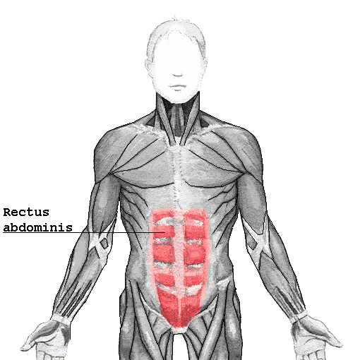
The rectus abdominis muscle, also known as the "abdominals" or "abs", is a paired muscle running vertically on each side of the anterior wall of the human abdomen, as well as that of some other mammals. There are two parallel muscles, separated by a midline band of connective tissue called the linea alba. It extends from the pubic symphysis, pubic crest and pubic tubercle inferiorly, to the xiphoid process and costal cartilages of ribs V to VII superiorly. The proximal attachments are the pubic crest and the pubic symphysis. It attaches distally at the costal cartilages of ribs 5-7 and the xiphoid process of the sternum.
Contents
The rectus abdominis muscle is contained in the rectus sheath, which consists of the aponeuroses of the lateral abdominal muscles. Bands of connective tissue called the tendinous intersections traverse the rectus abdominus, which separates this parallel muscle into distinct muscle bellies. The outer, most lateral line, defining the "abs" is the linea semilunaris. In the abdomens of people with low body fat, these bellies can be viewed externally and are commonly referred to as "four", "six", "eight", or even "ten packs", depending on how many are visible; although six is the most common.
Structure
The rectus abdominis is a long flat muscle, which extends along the whole length of the front of the abdomen, and is separated from its fellow of the opposite side by the linea alba.
The upper portion, attached principally to the cartilage of the fifth rib, usually has some fibers of insertion into the anterior extremity of the rib itself.
Size
It's typically around 10 mm thick (compared to 20 mm thick superficial fat), or 20 mm thick in young athletes such as handball players. Typical volume is around 300 cm³ in non-active individuals, or almost 500 cm³ in athletes (tennis players).
Blood supply
The rectus abdominis has many sources of arterial blood supply. In reconstructive surgery terms, it is a Mathes and Nahai Type III muscle with two dominant pedicles. First, the inferior epigastric artery and vein (or veins) run superiorly on the posterior surface of the rectus abdominis, enter the rectus fascia at the arcuate line, and serve the lower part of the muscle. Second, the superior epigastric artery, a terminal branch of the internal thoracic artery, supplies blood to the upper portion. Finally, numerous small segmental contributions come from the lower six intercostal arteries as well.
Nerve supply
The muscles are innervated by thoraco-abdominal nerves, these are continuations of the T7-T11 intercostal nerves and pierce the anterior layer of the rectus sheath. Sensory supply is from the 7-12 thoracic nerves
Variation
The sternalis muscle may be a variant form of the pectoralis major or the rectus abdominis. Some fibers are occasionally connected with the costoxiphoid ligaments, and the side of the xiphoid process.
Function
The rectus abdominis is an important postural muscle. It is responsible for flexing the lumbar spine, as when doing a so-called "crunch" sit up. The rib cage is brought up to where the pelvis is when the pelvis is fixed, or the pelvis can be brought towards the rib cage (posterior pelvic tilt) when the rib cage is fixed, such as in a leg-hip raise. The two can also be brought together simultaneously when neither is fixed in space.
The rectus abdominis assists with breathing and plays an important role in respiration when forcefully exhaling, as seen after exercise as well as in conditions where exhalation is difficult such as emphysema. It also helps in keeping the internal organs intact and in creating intra-abdominal pressure, such as when exercising or lifting heavy weights, during forceful defecation or parturition (childbirth).
Clinical significance
An abdominal muscle strain, also called a pulled abdominal muscle, is an injury to one of the muscles of the abdominal wall. A muscle strain occurs when the muscle is stretched too far. When this occurs the muscle fibers are torn. Most commonly, a strain causes microscopic tears within the muscle, but occasionally, in severe injuries, the muscle can rupture from its attachment.
Other animals
The rectus abdominis is similar in most vertebrates. The most obvious difference between animal and human abdominal musculature is that in animals, there are a different number of tendinous intersections.