
Order Incertae sedis Rank Genus | ||
 | ||
Similar Iridoviridae, Iridovirus, Batrachochytrium dendrobatidis, Infectious hematopoietic necrosis, Tiger salamander | ||
Ranavirus amphibians
Ranavirus is a genus of viruses, in the family Iridoviridae. There are four other genera of viruses within the family Iridoviridae, but Ranavirus is the only one that includes viruses that are infectious to amphibians and reptiles. Additionally, it is one of the three genera within this family which infect teleost fishes, along with Lymphocystivirus and Megalocytivirus. The family Iridoviridae is one of the five families of nucleocytoplasmic large DNA viruses.
Contents
- Ranavirus amphibians
- Reptiles amphibians in us succumbing to deadly ranavirus
- Ecological impact
- Etymology
- Evolution
- Hosts
- Taxonomy
- Structure
- Replication
- Transmission
- Epizoology
- Pathogenesis
- Gross pathology
- References
Reptiles amphibians in us succumbing to deadly ranavirus
Ecological impact

The Ranaviruses, like the Megalocytiviruses, are an emerging group of closely related dsDNA viruses which cause systemic infections in a wide variety of wild and cultured fresh and saltwater fishes. As with Megalocytiviruses, Ranavirus outbreaks are therefore of considerable economic importance in aquaculture, as epizootics can result in moderate fish loss or mass mortality events of cultured fishes. Unlike Megalocytiviruses, however, Ranavirus infections in amphibians have been implicated as a contributing factor in the global decline of amphibian populations. The impact of Ranaviruses on amphibian populations has been compared to the chytrid fungus Batrachochytrium dendrobatidis, the causative agent of chytridiomycosis.
Etymology
Rana is derived from the Latin for "frog", reflecting the first isolation of a Ranavirus in 1960s from the Northern leopard frog (Lithobates pipiens).
Evolution
The ranaviruses appear to have evolved from a fish virus which subsequently infected amphibians and reptiles.
Hosts
Several reptile species are known to be affected:

Taxonomy
Group: dsDNA

The family Iridoviridae is divided into five genera which include Chloriridovirus, Iridovirus, Lymphocystivirus, Megalocytivirus, and Ranavirus. The genus Ranavirus is composed of 6 recognized viral species, 3 of which are known to infect amphibians (Ambystoma tigrinum virus (ATV), Bohle iridovirus (BIV), and frog virus 3).
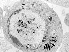
Structure
Ranaviruses are large icosahedral DNA viruses measuring approximately 150 nm in diameter with a large single linear dsDNA genome of roughly 105 kbp which codes for around 100 gene products. The main structural component of the protein capsid is the major capsid protein (MCP).
Replication
Ranaviral replication is well-studied using the type species for the genus, frog virus 3 (FV3). Replication of FV3 occurs between 12 and 32 degrees Celsius. Ranaviruses enter the host cell by receptor-mediated endocytosis. Viral particles are uncoated and subsequently move into the cell nucleus, where viral DNA replication begins via a virally encoded DNA polymerase. Viral DNA then abandons the cell nucleus and begins the second stage of DNA replication in the cytoplasm, ultimately forming DNA concatemers. The viral DNA is then packaged via a headful mechanism into infectious virions. The ranavirus genome, like other iridoviral genomes is circularly permuted and exhibits terminally redundant DNA.
Transmission
Transmission of ranaviruses is thought to occur by multiple routes, including contaminated soil, direct contact, waterborne exposure, and ingestion of infected tissues during predation, necrophagy or cannibalism. Ranaviruses are relatively stable in aquatic environments, persisting several weeks or longer outside a host organism.
Epizoology
Amphibian mass mortality events due to Ranavirus have been reported in Asia, Europe, North America, and South America. Ranaviruses have been isolated from wild populations of amphibians in Australia, but have not been associated with mass mortality on that continent.
Pathogenesis
Synthesis of viral proteins begins within hours of viral entry with necrosis or apoptosis occurring as early as a few hours post-infection.
Gross pathology
Gross lesions associated with Ranavirus infection include erythema, generalized swelling, hemorrhage, limb swelling, and swollen and friable livers.