
Dorlands/Elsevier r_07/12700361 FMA 57778 | TA A01.2.02.009 | |
 | ||
Latin Trigonum cervicale posteriusTrigonum colli posteriusRegio cervicalis posterior | ||
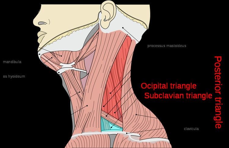
The posterior triangle (or lateral cervical region) is a region of the neck.
Contents
Boundaries
It has the following boundaries:
Apex: Union of the sternocleidomastoid and the trapezius muscles at the superior nuchal line of the occipital bone
Anterior: Posterior border of the sternocleidomastoideus
Posterior: Anterior border of the trapezius
Base: Middle one third of the clavicle
Roof: Investing layer of the deep cervical fascia
Divisions
The posterior triangle is crossed, about 2.5 cm above the clavicle, by the inferior belly of the omohyoid muscle, which divides the space into two triangles:
Contents
A) Nerves and Plexuses:
B) Vessels:
C) Lymph Nodes:
D) Muscles:
Clinical significance
The accessory nerve (CN XI) is particularly vulnerable to damage during lymph node biopsy. Damage results in an inability to shrug the shoulders or raise the arm above the head (e.g., for brushing hair), particularly due to compromised trapezius muscle innervation.
The external jugular vein's superficial location within the posterior triangle also makes it vulnerable to injury. It is also the site of clinical examination of Jugular venous pressure.
The right atrial pressure is reflected in it because there are no valve in the entire course of this vein and is straight, therefore used to examine Jugular Venous Pressure.
As external jugular vein pierces the fascia, the margins of the vein get adherent to fascia. So if the vein gets cut, it cannot close and air is sucked in due to negative intrathoracic pressure causes air embolism. To prevent this, fascia colli has to be cut.