
 | ||
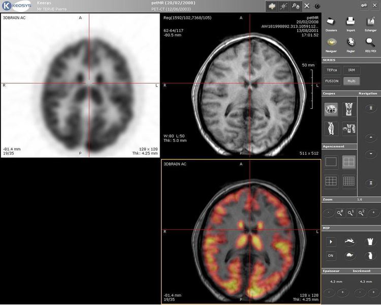
Positron emission tomography–magnetic resonance imaging (PET-MRI) is a hybrid imaging technology that incorporates magnetic resonance imaging (MRI) soft tissue morphological imaging and positron emission tomography (PET) functional imaging.
Contents
Applications
Presently, the main clinical fields of PET-MRI are oncology, cardiology and neurology. Research studies are actively conducted at the moment to understand benefits of the new PET-MRI diagnostic method. The technology combines the exquisite structural and functional characterization of tissue provided by MRI with the extreme sensitivity of PET imaging of metabolism and tracking of uniquely labeled cell types or cell receptors. There is discussion and investigation into utilizing PET-MR with Ion Therapy for the purpose of cancer treatment. with MRI's ability to accurately depict the proton density of tissue is a good match for the benefits and technical challenges of treatment planning utilizing Ion Therapy systems.
Manufacturers
Currently four companies offer combined PET-MR systems: Philips, Siemens, GE, Aspect Imaging and MR Solutions. The first two clinical whole body PET-MRI systems were installed by Philips at Mount Sinai Medical Centre in the United States and at Geneva University Hospital in Europe in 2010. One company, Cubresa, offers an MR-compatible preclinical PET scanner called NuPET™ for use in the bore of an existing MRI, enabling simultaneous PET/MR image acquisition. The first instrument was installed at the Izaak Walton Killam Health Centre in Halifax, Canada in 2016.
Clinical systems
Currently Siemens and GE are the only companies to offer a fully integrated whole body and simultaneous acquisition PET-MRI system. The Siemens system (Biograph mMR) received a CE mark and FDA approval for customer purchase in 2011.
The GE system (SIGNA PET/MR) received its 510K & CE mark in 2014.
The first fully RoHS compliant system was delivered in 2014. Over sixty facilities have since installed this technology.
Preclinical systems
Currently, the combination of positron emission tomography (PET) and magnetic resonance imaging (MRI) as a hybrid imaging modality is receiving great attention not only in its emerging clinical applications but also in the preclinical field. Several designs based on several different types of PET detector technology have been developed in recent years, some of which have been used for first preclinical studies.
A preclinical PET-MRI system with sequential acquisition is commercially available from Mediso Medical Imaging Systems since 2011. The first nanoScan PET-MRI system was installed at Karolinska Experimental Research and Imaging Centre of Karolinska Institutet in March 2011. The compact imaging system utilizes magnetically shielded position sensitive photomultiplier tubes and a compact 1 Tesla permanent magnet MRI platform. The integration has no adverse effects on the PET or on the MRI performance due to increased magnetic shielding of the PET component and low fringe field of the MRI component based on the performance evaluation of the system. Bruker Biospin, in collaboration with the University of Tübingen, have produced commercial prototype systems for their preclinical MRI magnets utilizing a Siemens PET platform. The first such system comprising a 7T Clinscan MRI with the PET insert was installed at the CAI in Brisbane Australia in 2012.
Aspect Imaging has collaborated with Brightonix Imaging from Korea and launched a simultaneous PET MRI for mice in 2016. The Aspect MRI system is based on a wide bore M7 1-Tesla permanent magnet.
MR Solutions’ high field, cryogen-free MRI system with an in-line PET module was installed at the University of Michigan in January 2016 and at the Georges-François Leclerc center in Dijon, France in February 2016. Data is acquired sequentially and co-registered together automatically.
The aforementioned NuPET™ MR-compatible preclinical scanner from Cubresa is designed to fit into a variety of existing MRI magnets, including those originally used for clinical imaging. Uniquely, this system enables simultaneous PET/MRI acquisition, allowing researchers to generate structural, functional, and molecular data that is both partially and temporally registered and acquired under identical physiological conditions. It can also be used as a compact, standalone PET scanner.
Hybrid PET MRI systems require special devices that balance tradeoffs between PET attenuation and MRI performance.
PET-MRI versus PET-CT
Comparisons have been made between PET-MRI and PET-CT, some sources stating that PET-MRI is simply an X-ray radiation-free version of PET-CT (PET-MRI has as well Radiation from Biomarker). In reality, there are differences beyond X-ray radiation dose.
This article written at the National Institute of Health in Bethesda, Maryland provides very good information on the technical differences between CT, MRI and PET and their combinations. It also references the advantages of simultaneous or single unit sequential over traditional sequential.