
Specialty Dentistry DiseasesDB 29362 MeSH D010518 | ICD-10 K05.4 MedlinePlus 001059 | |
 | ||
Pronunciation Periodontitis /ˌpɛrioʊdɒnˈtaɪtɪs/, pyorrhea /ˌpaɪəˈriə/ | ||
Periodontitis, also known as gum disease and pyorrhea, is a set of inflammatory diseases affecting the tissues surrounding the teeth. Periodontitis involves progressive loss of the alveolar bone around the teeth, and if left untreated, can lead to the loosening and subsequent loss of teeth.
Contents
- Classification
- Extent
- Severity
- Signs and symptoms
- Associated conditions
- Causes
- Mechanism
- Prevention
- Management
- Initial therapy
- Reevaluation
- Surgery
- Maintenance
- Alternative treatments
- Prognosis
- Epidemiology
- Etymology
- Economics
- Other animals
- References
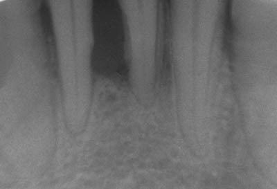
Periodontitis is caused by microorganisms that adhere to and grow on the tooth's surfaces, along with an over-aggressive immune response against these microorganisms. A diagnosis of periodontitis is established by inspecting the soft gum tissues around the teeth with a probe (i.e., a clinical examination) and by evaluating the patient's X-ray films (i.e., a radiographic examination), to determine the amount of bone loss around the teeth. Specialists in the treatment of periodontitis are periodontists; their field is known as "periodontology" or "periodontics".
Classification
The 1999 classification system for periodontal diseases and conditions listed seven major categories of periodontal diseases, of which 2–6 are termed destructive periodontal disease, because the damage is essentially irreversible. The seven categories are as follows:
- Gingivitis
- Chronic periodontitis
- Aggressive periodontitis
- Periodontitis as a manifestation of systemic disease
- Necrotizing ulcerative gingivitis/periodontitis
- Abscesses of the periodontium
- Combined periodontic-endodontic lesions
Moreover, terminology expressing both the extent and severity of periodontal diseases are appended to the terms above to denote the specific diagnosis of a particular patient or group of patients.
Extent
The "extent" of disease refers to the proportion of the dentition affected by the disease in terms of percentage of sites. Sites are defined as the positions at which probing measurements are taken around each tooth and, generally, six probing sites around each tooth are recorded, as follows:
- mesiobuccal
- mid-buccal
- distobuccal
- mesiolingual
- mid-lingual
- distolingual
If up to 30% of sites in the mouth are affected, the manifestation is classified as "localized"; for more than 30%, the term "generalized" is used.
Severity
The "severity" of disease refers to the amount of periodontal ligament fibers that have been lost, termed "clinical attachment loss". According to the American Academy of Periodontology, the classification of severity is as follows:
Signs and symptoms
In the early stages, periodontitis has very few symptoms, and in many individuals the disease has progressed significantly before they seek treatment.
Symptoms may include:
Patients should realize gingival inflammation and bone destruction are largely painless. Hence, people may wrongly assume painless bleeding after teeth cleaning is insignificant, although this may be a symptom of progressing periodontitis in that patient.
Associated conditions
Periodontitis has been linked to increased inflammation in the body, such as indicated by raised levels of C-reactive protein and interleukin-6. It is linked through this to increased risk of stroke, myocardial infarction, and atherosclerosis. It also linked in those over 60 years of age to impairments in delayed memory and calculation abilities. Individuals with impaired fasting glucose and diabetes mellitus have higher degrees of periodontal inflammation, and often have difficulties with balancing their blood glucose level owing to the constant systemic inflammatory state, caused by the periodontal inflammation. Although no causal association was proven, a recent study showed correlation between chronic periodontitis and erectile dysfunction.
Causes
Periodontitis is an inflammation of the periodontium, i.e., the tissues that support the teeth. The periodontium consists of four tissues:
The primary cause of gingivitis is poor or ineffective oral hygiene, which leads to the accumulation of a mycotic and bacterial matrix at the gum line, called dental plaque. Other contributors are poor nutrition and underlying medical issues such as diabetes. Diabetics must be meticulous with their homecare to control periodontal disease. New finger prick tests have been approved by the Food and Drug Administration in the US, and are being used in dental offices to identify and screen patients for possible contributory causes of gum disease, such as diabetes.
In some people, gingivitis progresses to periodontitis – with the destruction of the gingival fibers, the gum tissues separate from the tooth and deepened sulcus, called a periodontal pocket. Subgingival microorganisms (those that exist under the gum line) colonize the periodontal pockets and cause further inflammation in the gum tissues and progressive bone loss. Examples of secondary causes are those things that, by definition, cause microbic plaque accumulation, such as restoration overhangs and root proximity.
Smoking is another factor that increases the occurrence of periodontitis, directly or indirectly, and may interfere with or adversely affect its treatment.
Ehlers–Danlos syndrome is a periodontitis risk factor and so is the Papillon–Lefèvre syndrome also known as palmoplantar keratoderma.
If left undisturbed, microbial plaque calcifies to form calculus, which is commonly called tartar. Calculus above and below the gum line must be removed completely by the dental hygienist or dentist to treat gingivitis and periodontitis. Although the primary cause of both gingivitis and periodontitis is the microbial plaque that adheres to the tooth surfaces, there are many other modifying factors. A very strong risk factor is one's genetic susceptibility. Several conditions and diseases, including Down syndrome, diabetes, and other diseases that affect one's resistance to infection, also increase susceptibility to periodontitis.
Another factor that makes periodontitis a difficult disease to study is that human host response can also affect the alveolar bone resorption. Host response to the bacterial-mycotic insult is mainly determined by genetics; however, immune development may play some role in susceptibility.
According to some researchers periodontitis may be associated with higher stress. Periodontitis occurs more often in people from the lower end of the socioeconomic scale than people from the upper end of the socioeconomic scale.
Mechanism
As dental plaque or biofilm accumulates on the teeth near and below the gums, there is a shift in the composition of the biofilm from essentially streptococcus to an actinomyces dominant plaque. Motile bacteria is also seen more frequently. As this happens, inflammation sets in the gingiva. Initially, this takes the form of gingivitis, which represents inflammation confined to the soft tissues above the bone level. Inflammation in the gingiva can remain at the gingivitis level for a long period and will not progress to periodontitis, unless in the presence of local conditions or generalized host susceptibility. When this shift occurs, the immune system's response to plaque accumulation shifts from a predominantly neutrophilic mediated response to lymphocytic and plasma cell-mediated response. Clinically, the gingiva presents swelling, redness and a tendency to bleed. This modifies the environment, leading to changes in the composition of the biofilm itself. As this happens, a predominantly gram-negative environment is established, with periodontal pathogens emerging. These include A. actinomycetemcomitans, the red complex bacteria (P. gingivalis, T. Forsythia, T denticola) and to a lesser extent the orange complex bacteria (F nucleatum, P micros, P.intermedia, P. nigrecens, E. nodatum and S. constellates). Strongest bacterial association to chronic periodontitis is with P. Gingivalis. Numerous virulence factors have been identified for this pathogen. This allows P. gingivalis to elude defense mechanism and perpetuate inflammation inside the periodontium. Prolonged inflammation in the periodontium leads to an apical shift in the attachment of the gingiva to the tooth with deepening pockets and bone loss around the teeth. Untreated periodontitis progresses unevenly over time but results in loss of function, tissue destruction, and tooth loss.
Prevention
Daily oral hygiene measures to prevent periodontal disease include:
Typically, dental hygienists (or dentists) use special instruments to clean (debride) teeth below the gumline and disrupt any plaque growing below the gumline. This is a standard treatment to prevent any further progress of established periodontitis. Studies show that after such a professional cleaning (periodontal debridement), microbial plaque tends to grow back to precleaning levels after about three to four months. Nonetheless, the continued stabilization of a patient's periodontal state depends largely, if not primarily, on the patient's oral hygiene at home, as well as on the go. Without daily oral hygiene, periodontal disease will not be overcome, especially if the patient has a history of extensive periodontal disease.
Periodontal disease and tooth loss are associated with an increased risk, in male patients, of cancer.
Contributing causes may be high alcohol consumption or a diet low in antioxidants.
Management
The cornerstone of successful periodontal treatment starts with establishing excellent oral hygiene. This includes twice-daily brushing with daily flossing. Also, the use of an interdental brush is helpful if space between the teeth allows. For smaller spaces, products such as narrow picks with soft rubber bristles provide excellent manual cleaning. Persons with dexterity problems, such as arthritis, may find oral hygiene to be difficult and may require more frequent professional care and/or the use of a powered toothbrush. Persons with periodontitis must realize it is a chronic inflammatory disease and a lifelong regimen of excellent hygiene and professional maintenance care with a dentist/hygienist or periodontist is required to maintain affected teeth.
Initial therapy
Removal of microbial plaque and calculus is necessary to establish periodontal health. The first step in the treatment of periodontitis involves nonsurgical cleaning below the gumline with a procedure called scaling and debridement. In the past, root planing was used (removal of the cemental layer as well as calculus). This procedure involves the use of specialized curettes to mechanically remove plaque and calculus from below the gumline, and may require multiple visits and local anesthesia to adequately complete. In addition to initial scaling and root planing, it may also be necessary to adjust the occlusion (bite) to prevent excessive force on teeth that have reduced bone support. Also, it may be necessary to complete any other dental needs, such as replacement of rough, plaque-retentive restorations, closure of open contacts between teeth, and any other requirements diagnosed at the initial evaluation.
Reevaluation
Multiple clinical studies have shown nonsurgical scaling and root planing are usually successful if the periodontal pockets are shallower than 4–5 mm (0.16–0.20 in). The dentist or hygienist must perform a re-evaluation four to six weeks after the initial scaling and root planing, to determine if the patient's oral hygiene has improved and inflammation has regressed. Probing should be avoided then, and an analysis by gingival index should determine the presence or absence of inflammation. The monthly reevaluation of periodontal therapy should involve periodontal charting as a better indication of the success of treatment, and to see if other courses of treatment can be identified. Pocket depths of greater than 5–6 mm (0.20–0.24 in) which remain after initial therapy, with bleeding upon probing, indicate continued active disease and will very likely lead to further bone loss over time. This is especially true in molar tooth sites where furcations (areas between the roots) have been exposed.
Surgery
If nonsurgical therapy is found to have been unsuccessful in managing signs of disease activity, periodontal surgery may be needed to stop progressive bone loss and regenerate lost bone where possible. Many surgical approaches are used in the treatment of advanced periodontitis, including open flap debridement and osseous surgery, as well as guided tissue regeneration and bone grafting. The goal of periodontal surgery is access for definitive calculus removal and surgical management of bony irregularities which have resulted from the disease process to reduce pockets as much as possible. Long-term studies have shown, in moderate to advanced periodontitis, surgically treated cases often have less further breakdown over time and, when coupled with a regular post-treatment maintenance regimen, are successful in nearly halting tooth loss in nearly 85% of patients.
Maintenance
Once successful periodontal treatment has been completed, with or without surgery, an ongoing regimen of "periodontal maintenance" is required. This involves regular checkups and detailed cleanings every three months to prevent repopulation of periodontitis-causing microorganisms, and to closely monitor affected teeth so early treatment can be rendered if the disease recurs. Usually, periodontal disease exists due to poor plaque control, therefore if the brushing techniques are not modified, a periodontal recurrence is probable.
Alternative treatments
Periodontitis has an inescapable relationship with subgingival calculus (tartar). The first step in any procedure is to eliminate calculus under the gum line, as it houses destructive anaerobic microorganisms that consume bone, gum and cementum (connective tissue) for food.
Most alternative "at-home" gum disease treatments involve injecting antimicrobial solutions, such as hydrogen peroxide, into periodontal pockets via slender applicators or oral irrigators. This process disrupts anaerobic micro-organism colonies and is effective at reducing infections and inflammation when used daily. A number of other products, functionally equivalent to hydrogen peroxide, are commercially available, but at substantially higher cost. However, such treatments do not address calculus formations, and so are short-lived, as anaerobic microbial colonies quickly regenerate in and around calculus.
Doxycycline may be given alongside the primary therapy of scaling (see § initial therapy). Doxycycline has been shown to improve indicators of disease progression (namely probing depth and attachment level). Its mechanism of action involves inhibition of matrix metalloproteinases (such as collagenase), which degrade the teeth's supporting tissues (periodontium) under inflammatory conditions. To avoid killing beneficial oral microbes, only small doses of doxycycline (20 mg) are used.
Prognosis
Dentists and dental hygienists measure periodontal disease using a device called a periodontal probe. This thin "measuring stick" is gently placed into the space between the gums and the teeth, and slipped below the gumline. If the probe can slip more than 3 mm (0.12 in) below the gumline, the patient is said to have a gingival pocket if no migration of the epithelial attachment has occurred or a periodontal pocket if apical migration has occurred. This is somewhat of a misnomer, as any depth is, in essence, a pocket, which in turn is defined by its depth, i.e., a 2-mm pocket or a 6-mm pocket. However, pockets are generally accepted as self-cleansable (at home, by the patient, with a toothbrush) if they are 3 mm or less in depth. This is important because if a pocket is deeper than 3 mm around the tooth, at-home care will not be sufficient to cleanse the pocket, and professional care should be sought. When the pocket depths reach 6 to 7 mm (0.24 to 0.28 in) in depth, the hand instruments and cavitrons used by the dental professionals may not reach deeply enough into the pocket to clean out the microbial plaque that causes gingival inflammation. In such a situation, the bone or the gums around that tooth should be surgically altered or it will always have inflammation which will likely result in more bone loss around that tooth. An additional way to stop the inflammation would be for the patient to receive subgingival antibiotics (such as minocycline) or undergo some form of gingival surgery to access the depths of the pockets and perhaps even change the pocket depths so they become 3 mm or less in depth and can once again be properly cleaned by the patient at home with his or her toothbrush.
If patients have 7-mm or deeper pockets around their teeth, then they would likely risk eventual tooth loss over the years. If this periodontal condition is not identified and the patients remain unaware of the progressive nature of the disease, then years later, they may be surprised that some teeth will gradually become loose and may need to be extracted, sometimes due to a severe infection or even pain.
According to the Sri Lankan tea laborer study, in the absence of any oral hygiene activity, approximately 10% will suffer from severe periodontal disease with rapid loss of attachment (>2 mm/year). About 80% will suffer from moderate loss (1–2 mm/year) and the remaining 10% will not suffer any loss.
Epidemiology
Periodontitis is very common, and is widely regarded as the second most common dental disease worldwide, after dental decay, and in the United States has a prevalence of 30–50% of the population, but only about 10% have severe forms.
Chronic periodontitis affects about 750 million people or about 10.8% of the population as of 2010.
Like other conditions intimately related to access to hygiene and basic medical monitoring and care, periodontitis tends to be more common in economically disadvantaged populations or regions. Its occurrence decreases with a higher standard of living. In Israeli population, individuals of Yemenite, North-African, South Asian, or Mediterranean origin have higher prevalence of periodontal disease than individuals from European descent. Periodontitis is frequently reported to be socially patterned, i.e. people from the lower end of the socioeconomic scale suffer more often from it than people from the upper end of the socioeconomic scale.
Etymology
The word "periodontitis" (Greek: περιοδοντίτις) comes from the Greek peri, "around", odous (GEN odontos), "tooth", and the suffix -itis, in medical terminology "inflammation". The word pyorrhea (alternative spelling: pyorrhoea) comes from the Greek pyorrhoia (πυόρροια), "discharge of matter", itself from pyon, "discharge from a sore", rhoē, "flow", and the suffix -ia. In English this term can describe, as in Greek, any discharge of pus; i.e. it is not restricted to these diseases of the teeth.
Economics
It is estimated that periodontitis results in worldwide productivity losses in the size of about US$54 billion yearly.
Other animals
Periodontal disease is the most common disease found in dogs and affects more than 80% of dogs aged three years or older. Its prevalence in dogs increases with age, but decreases with increasing body weight; i.e., toy and miniature breeds are more severely affected. Recent research undertaken at the Waltham Centre for Pet Nutrition has established that the bacteria associated with gum disease in dogs are not the same as in humans. Systemic disease may develop because the gums are very vascular (have a good blood supply). The blood stream carries these anaerobic micro-organisms, and they are filtered out by the kidneys and liver, where they may colonize and create microabscesses. The microorganisms traveling through the blood may also attach to the heart valves, causing vegetative infective endocarditis (infected heart valves). Additional diseases that may result from periodontitis include chronic bronchitis and pulmonary fibrosis.