
 | ||
Many mammalian species have developed keratinized penile spines along the glans and/or shaft, which may be involved in sexual selection. These spines have been described as being simple, single-pointed structures (macaques) or complex with two or three points per spine (strepsirrhines). Penile spine morphology may be related to mating system.
Contents
Non-human mammals
Felines, especially domestic cats, are well known for having penile spines. Upon withdrawal of a cat's penis, the spines rake the walls of the female's vagina, which may cause ovulation.
Penile spines in chimpanzees and mice are small surface projections made by the piling up of keratinized cell layers in the outermost skin surface. They also occur in spotted hyenas, fossas, echidnas, bats, and several rodent species.
Humans
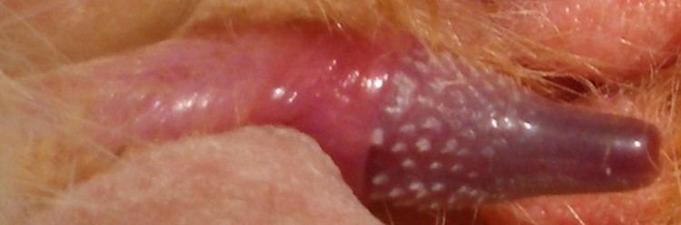
In contrast to chimpanzees and mice, a common morphological variant found in humans called Hirsuties coronae glandis, or pearly penile papules, are substantially larger, appear to be an outpocketing of both surface and underlying connective tissue layers, and lack the rich innervation seen in other animals. Thus, the relationship between the structures is still uncertain.
In the primate line, a regulatory DNA sequence associated with the formation of small keratinized penile spines was lost. This simplification of penis anatomy may be associated with the sexual habits of humans. In some species which retain the full expression of penile spines, penile spines contribute to increased sexual sensation and quicker orgasms.
Hirsuties coronae glandis, found in humans, are sometimes described as vestigial remnants of penile spines.
An hCONDEL (highly conserved region of DNA that contains deletions in humans) located near the locus of the androgen receptor gene may be responsible for the loss of penile spines in humans.
Birds
The penises of some bird species feature spines and brush-like filaments.