
Dorlands/Elsevier r_10/12705378 | FMA 44676 | |
 | ||
Latin Rete patellare, anastomosis patellaris, rete articulare genus TA A12.2.16.040A12.2.16.041 | ||
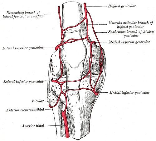
The patellar network (circulatory anastomosis around the knee-joint, patellar anastomosis, genicular anastomosis, articular vascular network of knee or rete articulare genus) is an intricate network of vessels around and above the patella, and on the contiguous ends of the femur and tibia, forming a superficial and a deep plexus.
The arteries which form this plexus are the inferior medial and superior medial genicular arteries, the inferior lateral and superior lateral genicular arteries, the descending genicular artery, the descending branch of lateral femoral circumflex artery, and the anterior tibial recurrent artery.
Clinical relevance
The genicular anastomosis provides collateral circulation to supply the leg when the knee is fully flexed.
When the knee suffers a popliteal aneurysm, if the femoral artery has to be ligated surgically, blood can still reach the popliteal artery distal to the ligation via the genicular anastomosis. However, if flow in the femoral artery of a normal leg is suddenly disrupted, blood flow distally is rarely sufficient. The reason for this is the fact that the genicular anastomosis is only present in a minority of individuals and is always undeveloped when disease in the femoral artery is absent.
Illustrations of the genicular anastomosis in textbooks all appear to have been derived from the idealized image, shown in the sidebox, produced first by Gray's Anatomy in 1910. Neither the 1910 illustration, nor any subsequent version, was made of an anatomical dissection but rather from the writings of John Hunter (surgeon) and Astley Cooper which described the genicular anastomosis many years after ligation of the femoral artery for popliteal aneurysm. The genicular anastomosis has not been demonstrated even with modern imaging techniques such as X-ray computed tomography or angiography.