
From patella MeSH A02.513.514.475 TA A03.6.08.015 | Latin ligamentum patellae Dorlands
/Elsevier l_09/12492768 | |
 | ||
To tuberosity of the tibia | ||
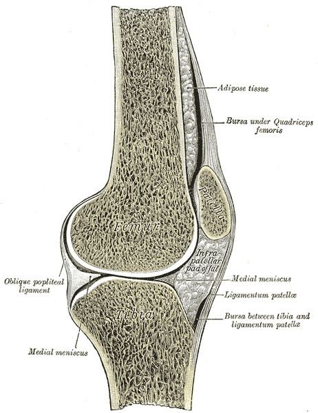
The patellar ligament is the distal portion of the common tendon of the quadriceps femoris, which is continued from the patella to the tibial tuberosity. It is also sometimes called the patellar tendon as it is a continuation of the quadriceps tendon.
Contents
Structure
The patellar ligament is a strong, flat, ligament, about 5 cm in length, which originates on the apex of the patella distally and adjoining margins of the patella and the rough depression on its posterior surface; below, it inserts on the tuberosity of the tibia; its superficial fibers are continuous over the front of the patella with those of the tendon of the quadriceps femoris.
The medial and lateral portions of the quadriceps tendon pass down on either side of the patella to be inserted into the upper extremity of the tibia on either side of the tuberosity; these portions merge into the capsule, as stated above, forming the medial and lateral patellar retinacula.
The posterior surface of the patellar ligament is separated from the synovial membrane of the joint by a large infrapatellar pad of fat, and from the tibia by a bursa.
Clinical significance
The patellar ligament can be injured in a patellar tendon rupture.
It can be used as a tissue source in the repair of other ligaments. In the event of a torn anterior cruciate ligament, the patellar ligament can be used in the rehabilitation process. In this case, the middle one third of the patellar ligament is harvested and inserted through tunnels that are drilled into the femur and tibia. The portion of the patellar ligament is then drawn through these tunnels in the bone and will be affixed to the bone via screws. The recovery process takes approximately 4–6 months upon the completion of surgery. This patellar ligament method of reconstruction was traditionally the gold standard graft for anterior cruciate ligament reconstruction, and is still one of the more preferred methods.
Its insertion on the tibia is the location of Osgood-Schlatter disease.