
Latin musculus papillaris TA A12.1.00.022 | Dorlands/Elsevier 12550081 FMA 76526 | |
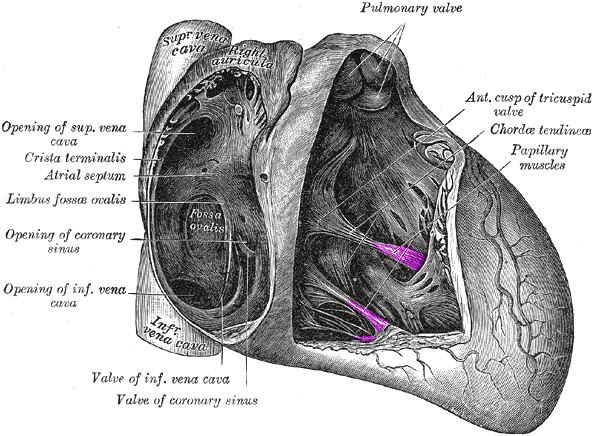 | ||
The papillary muscles are muscles located in the ventricles of the heart. They attach to the cusps of the atrioventricular valves (also known as the mitral and tricuspid valves) via the chordae tendineae and contract to prevent inversion or prolapse of these valves on systole (or ventricular contraction).
Contents
Structure
There are five total papillary muscles in the heart; three in the right ventricle and two in the left. The anterior, posterior, and septal papillary muscles of the right ventricle each attach via chordae tendineae to the tricuspid valve. The anterior and posterior papillary muscles of the left ventricle attach via chordae tendineae to the mitral valve.
Blood supply
The mitral valve papillary muscles in the left ventricle are called the anterolateral and posteromedial muscles.
The posteromedial muscle ruptures more frequently because it only has one source of blood supply.
Function
The papillary muscles of both the right and left ventricles begin to contract shortly before ventricular systole and maintain tension throughout. This prevents regurgitation—backward flow of ventricular blood into the atrial cavities—by bracing the atrioventricular valves against prolapse—being forced back into the atria by the high pressure in the ventricles.
Clinical significance
Papillary muscle rupture can be caused by a myocardial infarct, and dysfunction can be caused by ischemia. Both complications may lead to worsening of mitral regurgitation.