
Entrez 5209 | Ensembl ENSG00000170525 | |
 | ||
Aliases PFKFB3, IPFK2, PFK2, iPFK-2, 6-phosphofructo-2-kinase/fructose-2,6-biphosphatase 3 External IDs MGI: 2181202 HomoloGene: 88708 GeneCards: PFKFB3 | ||
PFKFB3 is a gene that encodes the 6-phosphofructo-2-kinase/fructose-2,6-biphosphatase 3 enzyme in humans. It is one of 4 tissue-specific PFKFB isoenzymes identified currently (PFKFB1-4).
Contents
Gene
The PFKFB3 gene is mapped to single locus on chromosome 10 (10p15-p14). It spans a region of 32.5kb with an open reading frame that is 5,675bp long. It is estimated to consist of 19 exons, of which 15 are regularly expressed. Alternative splicing of the variable, COOH-terminal domain has been observed, leading to 6 different isoforms termed UBI2K1 to UBI2K6 in humans. Different nomenclature also recognizes two broad categories of PFKFB3 isoforms, termed ‘inducible’ and ‘ubiquitous’. The inducible protein isoform, iPFK2, is named as such because its expression has been shown to be induced by hypoxic conditions.
The PFKFB3 promoter is predicted to contain multiple binding sites, including Sp-1 and AP-2 binding sites. It also contains motifs for the binding of E-box, nuclear factor-1 (NF-1), and progesterone response element. Expression of the promoter is shown to be induce by phorbol esters and cyclic-AMP-dependent protein kinase signaling.
Structure
The four PFKFB isoforms share high (85%) ‘2-Kase/2-Pase core’ sequence homology, but have different properties based on variable N- and C- terminal regulatory domains and variation in residues surrounding the surrounding the active sites. The PFKFB3 inducible isoform has higher ‘2-Kase’ (kinase) activity than other isoforms, due to phosphorylation of Ser-460 by PKA or AMP-dependent protein kinase. The high ‘2-Kase’ activity of PFKFB3 is also due to the lack of a specific Ser that is phosphorylated in the other PFKFB isoforms to decrease kinase activity.
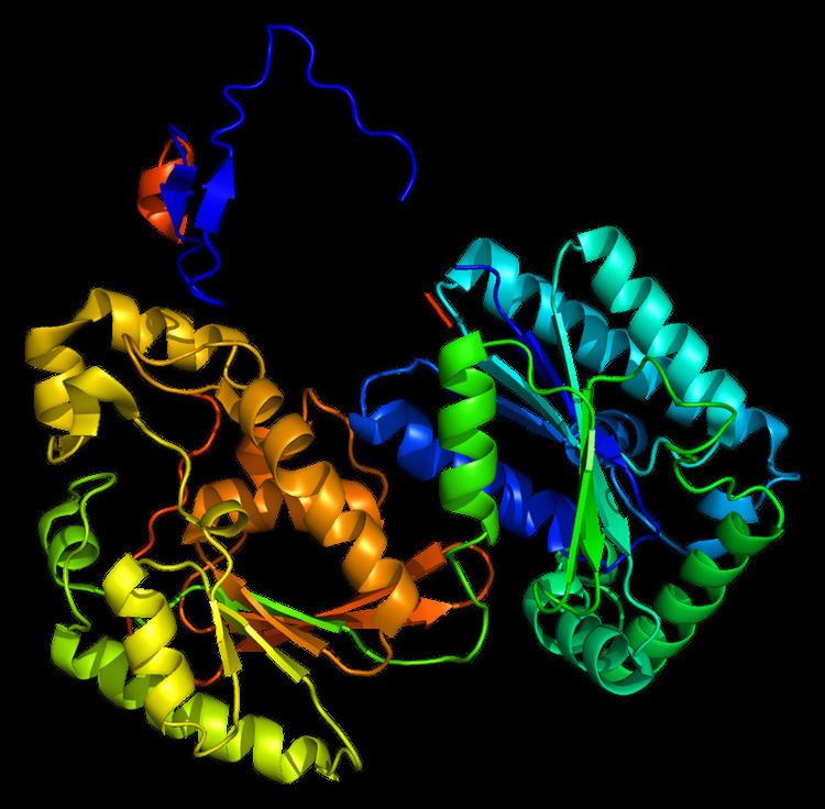
The primary protein encoded by PFKFB3, iPFK2, consists of 590 amino acids. It has a predicted molecular weight of 66.9 kDa and an isoelectric point of 8.64. The crystal structure was determined in 2006:
Function
iPFK2 converts fructose-6-phosphate to fructose-2,6-bisP (F2,6BP). F2,6BP is a ‘potent’ allosteric activator of 6-phosphofructokinase-1 (PFK-1), stimulating glycolysis. Click to see image of PFFKB3 function.
Warburg Effect
The Warburg effect, proposed by Otto Warbug in 1956, describes the upregulation of glycolysis in most cancer cells, even in the presence of oxygen. The high rate of glycolysis is accompanied by increased lactic acid fermentation, providing additional nutrients for cancer cell growth and tumorigenesis.
PFKFB3 is associated with the Warburg effect because its activity increases the rate of glycolysis. PFKFB3 has been found to be upregulated in numerous cancers, including colon, breast, ovarian, and thyroid. Reduced methylation of PFKFB3 is also found in some cancers, triggering the shift to the glycolytic pathway that supports cancerous growth.
Hypoxia Signaling Pathway
PFKFB3 expression is induced by hypoxia. The promoter of PFKFB3 contains binding sites, called hypoxia response elements (HREs), that recruit the binding of hypoxia-inducible factor-1 (HIF-1).
Hypoxia signaling via HIF-1α stabilization upregulates the transcription of genes that permit survival in low oxygen conditions. These genes include glycolysis enzymes, like PFKFB3, that permit ATP synthesis without oxygen, and vascular endothelial growth factor (VEGF), which promotes angiogenesis.
Cell Cycle & Apoptosis
It was more recently discovered that PFKFB3 promotes cell cycle progression (cell proliferation) and suppresses apoptosis by regulating cyclin-dependent kinase 1 (Cdk-1). PFKFB3’s synthesis of F2,6BP in the nucleus was found to regulate Cdk-1, whereas cytosolic PFKFB3 activates PFK-1. Nuclear PFKFB3 activates Cdk1 to phosphorylate the Thr-187 site of p27, causing decreased levels of p27. (See summary figure). Reduced p27 causes protection against apoptosis and progression of cells through the G1/S phase checkpoint These findings established a significant link between PFKFB3 cancer cell survival and proliferation.
Circadian Clock
Circadian clocks dysregulation is associated with many types of cancer. PFKFB3 expression exhibits circadian rhythmicity that is different between cancerous and non-cancerous cells. It was specifically found that the circadian-driven transcription factor ‘CLOCK’ binds to the PFKFB3 promoter at a genuine ‘E-box’ site to increase transcription in cancer cells.
Additional Cancer Connections
Anti-cancer Therapeutic Strategy
Inhibition of PFKFB3 is being analyzed as a potential anti-cancer therapy. Various small molecule inhibitors of PFKFB3 are currently in development. The most prominent inhibitor is 3PO, 3-(3-pyridinyl)-1-(4-pyridinyl)-2-propen-1-one. 3PO is a small molecule inhibitor that decreases glucose uptake and increases autophagy. Research is currently exploring various 3PO derivatives (ie. PFKF15) in an effort to increase their efficacy as anti-cancer therapies.
Autophagy
Enhanced activity of PFKFB3 accelerates ROS production as an end product of glycolysis, and thus increases autophagy. Likewise, inhibition of PFKFB3 has been found to induce autophagy. See summary image.
Autophagy can prolong cellular survival during low energy conditions. This finding was discovered in relation to rheumatoid arthritis. It was found that RA T cell fail to upregulate autophagy, and knockout experiments placed PFKFB3 as an upstream regulator of this process.
Insulin Signaling Pathway
PFKFB3 was identified in a kinome screen as a regulator of insulin/IGF-1. Suppression of PFKFB3 was found to decrease insulin-stimulated glucose uptake, GLUT4 translocation, and Akt signaling in 3T3-L1 adipocytes. Overexpression caused the insulin-dependent phosphorylation of Akt and Akt substrates.
PFKFB3 expression increases in fat tissues during adipogenesis, but prolonged insulin exposure has been shown to decrease the expression of PFKFB3. This is thought to occur due to a negative feedback mechanism involving insulin.
p38/MK2 Stress Sigaling Pathway
p38 MAPK have been found to increase PFKFB3 activity through (1) the transcriptional activation of PFKFB3 in response to stress stimuli and (2) the post-translational phosphorylation of iPFK2 at Ser-461.
See summary figure.