
Latin Septum orbitale TA A15.2.07.003 | Dorlands/Elsevier s_08/12730514 FMA 59328 | |
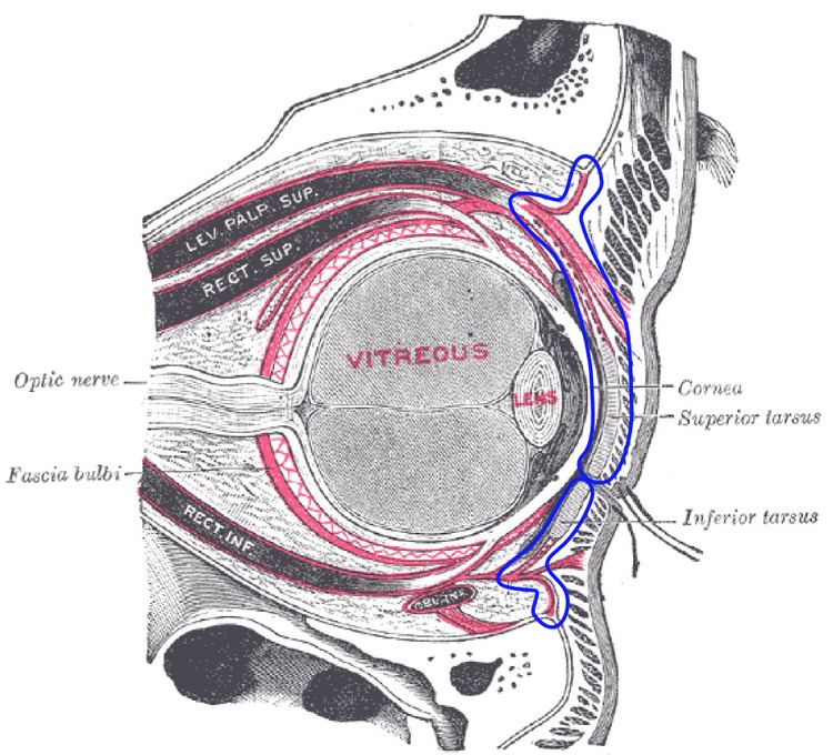 | ||
The orbital septum (Palpebral fascia) is a membranous sheet that acts as the anterior boundary of the orbit. It extends from the orbital rims to the eyelids. It forms the fibrous portion of the eyelids.
Contents
Anatomy
In the upper eyelid, the orbital septum blends with the tendon of the levator palpebrae superioris, and in the lower eyelid with the tarsal plate.
When the eyes are closed, the whole orbital opening is covered by the septum and tarsi. Medially it is thin, and, becoming separated from the medial palpebral ligament, attaches to the lacrimal bone at its posterior crest. The medial ligament and its much weaker lateral counterpart, attached to the septum and orbit, keep the lids stable as the eye moves.
The septum is perforated by the vessels and nerves which pass from the orbital cavity to the face and scalp.
Clinical significance
With age the septum may weaken, and as a result orbital fat may herniate forwards. The operation to correct this is called blepharoplasty.
The orbital septum is an important landmark in distinguishing between orbital cellulitis (inside the septum) and periorbital cellulitis (outside the septum).