
Specialty emergency medicine | ICD-10 S02.3 | |
 | ||
An orbital blowout fracture is a traumatic deformity of the orbital floor or medial wall, typically resulting from impact of a blunt object larger than the orbital aperture, or eye socket. There are two broad categories of blowout fractures: open door, which are large, displaced and comminuted, and trapdoor, which are linear, hinged, and minimally displaced. They are characterized by double vision, sunken ocular globes, and loss of sensation of the cheek and upper gums due to infraorbital nerve injury.
Contents
In pure orbital blowout fractures, the orbital rim (the most anterior bony margin of the orbit) is preserved, while with impure fractures, the orbital rim is also injured. With the trapdoor variant, there is a high frequency of extra-ocular muscle entrapment, despite minimal signs of external trauma, a phenomenon referred to as a 'white-eyed' orbital blowout fracture. They can occur with other injuries such as transfacial Le Fort fractures or zygomaticomaxillary complex fractures. The most common causes are assault and motor vehicle accidents. In children, the trapdoor subtype are more common. Reconstruction is usually performed with a titanium mesh or porous polyethylene through a transconjunctival or subciliary incision. More recently, there has been success with endoscopic, or minimally invasive, approaches.
History
Orbital floor fractures were investigated and described by MacKenzie in Paris in 1844 and the term blow out fracture was coined in 1957 by Smith & Regan, who were investigating injuries to the orbit and resultant inferior rectus entrapment, by placing a hurling ball on cadaverous orbits and striking it with a mallet.
Mechanism
The force of a blow to the orbit is dissipated by a fracture of the surrounding bone, usually the orbital floor and/or the medial orbital wall. Serious consequences of such injury include diplopia on upward gaze where there is significant damage to the orbital floor. In blowout fractures, the medial wall is fractured indirectly. When an external force is applied to the orbital cavity from an object whose diameter is larger than that of the orbit, the orbital contents are retropulsed and compressed. The consequent sudden rise in intraorbital pressure is transmitted to the walls of the orbit, which ultimately leads to fractures of the thin medial wall and/or orbital floor. Theoretically, this mechanism should lead to more fractures of the medial wall than the floor, since the medial wall is slightly thinner (0.25 mm vs 0.50 mm). However, it is known that pure blowout fractures most frequently involve the orbital floor. This may be attributed to the honeycomb structure of the numerous bony septa of the ethmoid sinuses, which support the lamina papyracea, thus allowing it to withstand the sudden rise in intraorbital hydraulic pressure better than the orbital floor.
Signs
Some clinically observed signs include:
Causes
Common medical causes of blowout fracture may include:
Diagnosis
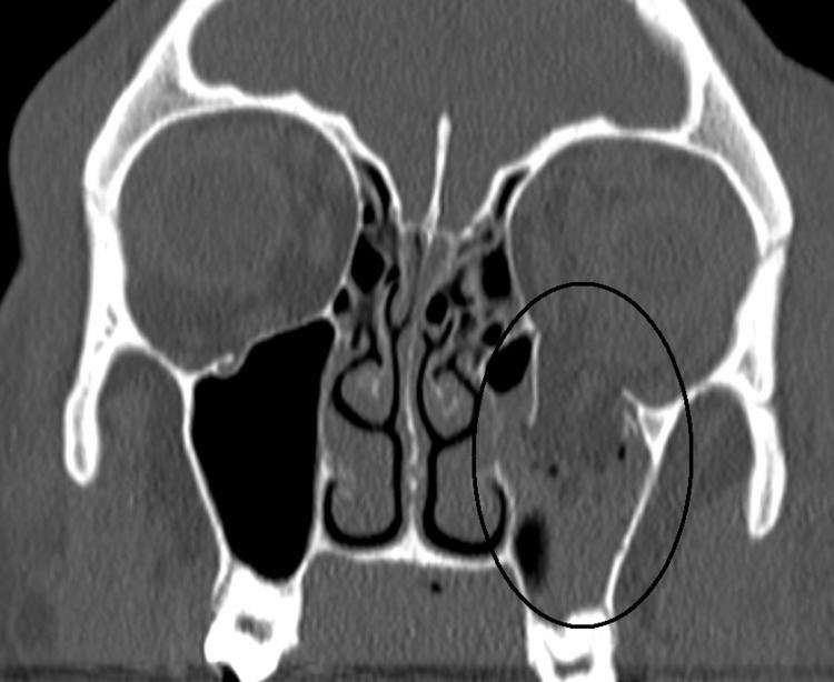
The most commonly fractured area in blowout fracture is the floor of orbit. Diagnosis is based on clinical and radiographic evidence. Periorbital bruising and subconjunctival hemorrhage are indirect signs of a possible fracture. On Water's view radiograph, polypoid mass can be observed hanging from the floor into the maxillary antrum, classically known as teardrop sign, as it usually is in shape of a teardrop. This polypoid mass consists of herniated orbital contents, periorbital fat and inferior rectus muscle. The affected sinus is partially opacified on radiograph. Air-fluid level in maxillary sinus may sometimes be seen due to presence of blood. CT scan can also show any soft tissue and bone involvement. Fracture of medial wall can produce subcutaneous emphysema, especially when blowing the nose or while sneezing. Lucency in orbits (on a radiograph) usually indicate orbital emphysema.
Treatment
Surgery is indicated if there is enophthalmos greater than 2 mm on imaging, Double vision on primary or inferior gaze, entrapment of extraocular muscles, or the fracture involves greater than 50% of the orbital floor. When not surgically repaired, most blowout fractures heal spontaneously without significant consequence. Corticosteroid therapy may be used to reduce swelling. Antibiotics are usually given as prevention of infection.
Surgical repair of a "blowout" is rarely undertaken immediately; it can be safely postponed for up to two weeks, if necessary, to let the swelling subside. Surgery to place an orbital implant leaves little or no scarring and the recovery period is usually brief. Hopefully, the surgery will provide a permanent cure, but sometimes it provides only partial relief from double vision or a sunken eye.