
NeuroLex ID Optic chiasm FMA 62045 | Latin chiasma opticum TA A14.1.08.403 | |
 | ||
MeSH A08.800.800.120.680.600 | ||
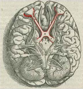
The optic chiasm or optic chiasma ( /ɒptɪk kaɪæzəm/; Greek χίασμα, "crossing", from the Greek χιάζω 'to mark with an X', after the Greek letter 'Χ', chi) is the part of the brain where the optic nerves partially cross. The optic chiasm is located at the bottom of the brain immediately below the hypothalamus. The optic chiasm is found in all vertebrates, although in cyclostomes (lampreys and hagfishes) it is located within the brain.
Contents
Structure
The optic nerve fibres on the nasal sides of each retina cross over (decussate) to the opposite side of the brain via the optic nerve at the optic chiasm (decussation of medial fibers). The temporal hemiretina, on the other hand, stays on the same side. The inferonasal retina are related to anterior portion of the optic chiasm whereas superonasal retinal fibers are related to the posterior portion of the optic chiasm.
The crossing over of optic nerve fibres at the optic chiasm allows the visual cortex to receive the same hemispheric visual field from both eyes. Superimposing and processing these monocular visual signals allow the visual cortex to generate binocular and stereoscopic vision. For example, the right visual cortex receives the nasal visual field from the left eye, and the temporal visual field from the right eye, which results in the right visual cortex producing a binocular image of the left hemispheric visual field. The net result of optic nerve crossing over at the optic chiasm is for the right cerebral hemisphere to sense and process left hemispheric vision, and for the left cerebral hemisphere to sense and process right hemispheric vision.
This crossing is an adaptive feature of frontally oriented eyes, found mostly in predatory animals requiring precise visual depth perception. (Prey animals, with laterally positioned eyes, have little binocular vision, so there is a more complete crossover of visual signals.) Beyond the optic chiasm, with crossed and uncrossed fibers, the optic nerves become optic tracts. The signals are passed on to the lateral geniculate body, in turn giving them to the occipital cortex (the outer matter of the rear brain).
Other animals
In Siamese cats with certain genotypes of the albino gene, this wiring is disrupted, with more of the nerve-crossing than is normal, as a number of scholars have reported. To compensate for lack of crossing in their brains, they cross their eyes (strabismus).
This is also seen in albino tigers, as Guillery & Kaas report.
History
The crossing of nerve fibres, and the impact on vision that this had, was probably first identified by Persian physician Zayn al-Din Gorgani (1042-1137).