
 | ||
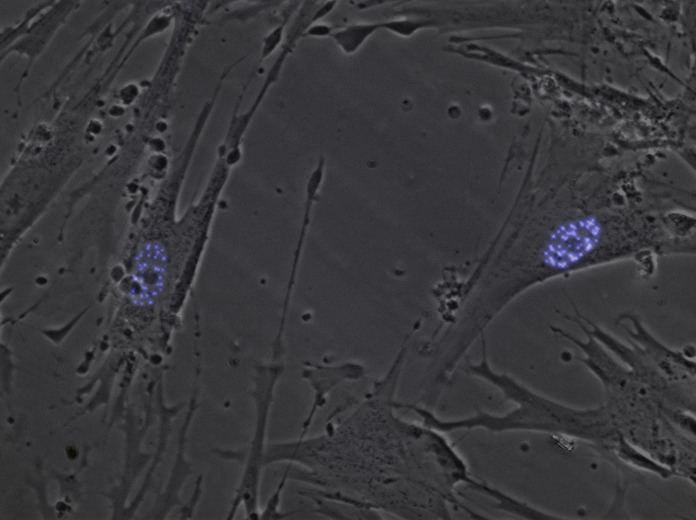
Nuclear dots (also known as Nuclear bodies, nuclear domains, or PML bodies) are punctate structures found in the nuclei of certain cells. Nuclear bodies (NBs) were first seen as prominent interchromatin structures in the nuclei of malignant or hyperstimulated animal cells identified using anti-sp100 autoantibodies from primary biliary cirrhosis and subsequently the promyelocytic leukemia (PML) factor, but appear also to be elevated in many autoimmune and cancerous diseases. Nuclear dots are metabolically stable and resistant to nuclease digestion and salt extraction.
Contents
Structure
Simple nuclear bodies (types I and II) and the shells of complex NB (types III, IVa and V) consist of a non-chromatinic fibrillar material which is most likely proteinaceous. That nuclear bodies co-isolated with the nuclear matrix, and were linked to the fibrogranular nuclear matrix component by projections from the surface of the nuclear bodies. The primary components of the nuclear dots are the proteins sp100 nuclear antigen, LYSP100(a homolog of sp100), ISG20, PML antigen, NDP55 and 53kDa protein associated with the nuclear matrix. Other proteins, such as PIC1/SUMO-1, which are associated with nuclear pore complex also associate with nuclear dots. The proteins can reorganize in the nucleus, by increasing number of dispersion in response to different stress (stimulation or heat shock, respectively).
Function
One of the nuclear dot proteins appears to be involved in transcriptional active regions. Expression of PML antigen and sp100 is responsive to interferons. Sp100 seems to have transcriptional transactivating properties. PML protein was reported to suppress growth and transformation, and specifically inhibits the infection of vesicular stomatitis virus (VSV) (a rhabdovirus) and influenza A virus, but not other types of viruses. The SUMO-1 ubiquitin like protein is responsible for modifying PML protein such that it is targeted to dots. whereas overexpression of PML results in programmed cell death.
One hypothesized function of the dots is as a 'nuclear dump' or 'storage depot'. The nuclear bodies may not all perform the same function. Sp140 associates with certain bodies and appears to be involved in transcriptional activation.
Pathology
These, or similar, bodies have been found increased in the presence of lymphoid cancers and SLE (lupus). They are also observed at higher frequencies in subacute sclerosing panencephalitis in these instances antibodies to measles shows expression and localization to the bodies.