
 | ||
The term mycangium (pl., mycangia) is used in biology for special structures on the body of an animal that are adapted for the transport of symbiotic fungi (usually in spore form). This is seen in many xylophagous insects (e.g. horntails and bark beetles), which apparently derive much of their nutrition from the digestion of various fungi that are growing amidst the wood fibers. In some cases, as in ambrosia beetles, the fungi are the sole food, and the excavations in the wood are simply to make a suitable microenvironment for the fungus to grow. In other cases (e.g., the southern pine beetle, Dendroctonus frontalis), wood tissue is the main food, and fungi weaken the defense response from host plant.
Contents
- Origin
- Function
- Mycangium in bark and ambrosia beetles
- Mycangium in woodwasp horntail
- Mycangium in lizard beetle
- Mycangium in ship timber beetle
- Mycangium in leaf rolling weevil
- Mycangium and symbiotic inoculum
- References
The mites, which have their own type of mycangium (for historical reasons, mite taxonomists use the term sporotheca), ride on or live next to the beetles.
Origin
These structures were first described by Helene Francke-Grosmann at 1956. Then Lekh R. Batra coined the word mycangia: modern Latin, from Greek myco 'fungus' + angeion 'vessel'.
Function
The most common function of mycangia is preserving and releasing symbiont. Usually the symbiotic inoculum in mycangia will provide great help to their vectors. They could help the vector adapt to the new environment or become nutrients of the vectors themselves and their descendants. Therefore, mycangia play an important role in protecting the inoculum from degradation and contamination. The structures of mycangia always look like a pouch or a container with caps or small opening that reduce the possibility of contaminants from outside. How mycangia release their inoculum is still unknown now.
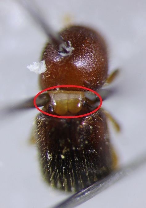
Mycangium in bark and ambrosia beetles
Mycangia of bark and ambrosia beetles (Curculionidae: Scolytinae and Platypodinae) are often complex cuticular invaginations for transport of symbiotic fungi. Phloem-feeding bark beetles (Coleoptera: Curculionidae: Scolytinae) have usually numerous small pits on the surface of their body, while ambrosia beetles (many Scolytinae and all Platypodinae), which are completely dependent on their fungal symbiont, have deep and complicated pouches. These mycangia are often equipped with glands secreting substances to support fungal spores and perhaps to nourish mycelium during transport. In many cases, the entrance to a mycangium is surrounded by tufts of setae, aiding in scraping mycelium and spores from walls of the tunnels and directing the spores into the mycangium. The mycangia in ambrosia beetle are highly diverse with different types in different genera or tribes. Some are oral mycangia, such as genus Ambrosiodmus (see left fig) and Euwallacea. Some are pronotal mycangia, such Xylosandrus and Cnestus.
Mycangium in woodwasp (horntail)
Mycangia of the woodwasp (Hymenoptera: Siricidae) were first described by Buchner. Different from highly diverse types in bark and ambrosia beetles, woodwasps only have a pair of mycangia on the top of their ovipositor. Then when females deposit their eggs inside the host plant, they inject the symbiotic fungi from mycangia and phytotoxic mucus from another reservoir-like structure.
Mycangium in lizard beetle
One species of lizard beetle Doubledaya bucculenta (Coleoptera: Erotylidae: Erotylidae) has mycangia on the tergum of the eighth abdominal segment. This ovipositor-associated mycangia is only present in adult females. Before Doubledaya bucculentnta deposit their eggs and inject the symbiotic microorganisms on a recently dead bamboo, they will excavate a small hole through the bamboo culm.
Mycangium in ship-timber beetle
The ship-timber beetle (Coleoptera: Lymexylidae) is another family of wood-boring beetles that live with symbiotic fungi. Buchner first discovered their mycangia located in the ventral side of the long ovipositor. These mycangia form a pair of integumental pouches at either side near the tip of oviduct. When the female lays the eggs, new eggs are coated with the fungal spores.
Mycangium in leaf-rolling weevil
Females of the leaf-rolling weevil in the genus Euops (Coleoptera: Attelabidae) store symbiotic fungi in the mycangia, which is between the first ventral segment of the abdomen and the thorax. Different from ovipositor-associate mycangia in woodwasps, lizard beetles, and ship-timber beetles, mycangia of leaf-rolling weevils is a pair of spore incubators at the anterior end of the abdomen. This mycangium is formed by the coxa and the metendosternite at the posterior end of the thorax.
Mycangium and symbiotic inoculum
Most of the inoculum in mycangia are fungi. The symbiotic inoculum of most bark and ambrosia beetles are fungi belonging to Ophiostomatales (Ascomycota: Sordariomycetidae) and Microascales (Ascomycota: Hypocreomycetidae). Symbiotic fungi in mycangia of woodwasps are Amylostereaceae (Basidiomycota: Russulales). Symbiotic fungi in mycangia of lizard beetles are yeast (Ascomycota: Saccharomycetales). Symbiotic fungi in mycangia of ship-timber beetles are Endomyces (Ascomycota: Dipodascaceae). Symbiotic fungi in mycangia of leaf-rolling weevils are Penicillium fungi (Ascomycota: Trichocomaceae). In addition to the above primary symbiotic fungi, secondary fungi and some Bacteria have been isolated from mycangia.