
Latin fusus neuromuscularis FMA 83607 | Code TH H3.11.06.0.00018 | |
 | ||
Muscle spindles are sensory receptors within the belly of a muscle that primarily detect changes in the length of this muscle. They convey length information to the central nervous system via sensory neurons. This information can be processed by the brain to determine the position of body parts. The responses of muscle spindles to changes in length also play an important role in regulating the contraction of muscles, by activating motor neurons via the stretch reflex to resist muscle stretch.
Contents
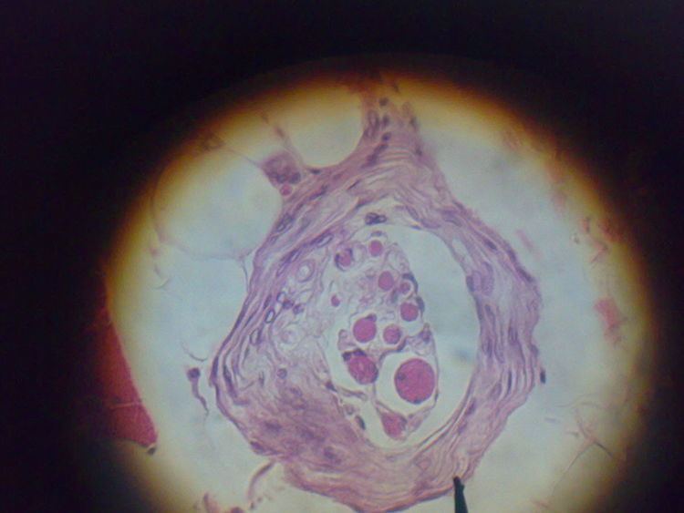
Muscle spindles are found within the belly of muscles, embedded in extrafusal muscle fibers. Note that "fusus" is the Latin word for spindle. Muscle spindles are composed of 3-12 intrafusal muscle fibers, of which there are three types:
Axons of gamma motoneurons also terminate in muscle spindles; they make synapses at either or both of the ends of the intrafusal muscle fibers and regulate the sensitivity of the sensory afferents, which are located in the non-contractile central (equatorial) region.
Muscle spindles are encapsulated by connective tissue, and are aligned parallel to extrafusal muscle fibers, unlike Golgi tendon organs, which are oriented in series.
The muscle spindle has both sensory and motor components.
Fusimotor neurons are classified as static or dynamic according to the type of intrafusal muscle fibers they innervate and their physiological effects on the responses of the Ia and II sensory neurons innervating the central, non-contractile part of the muscle spindle.
Sensitivity modification
The function of the gamma motoneurons is not to supplement the force of muscle contraction provided by the extrafusal fibers, but to modify the sensitivity of the muscle spindle sensory afferents to stretch. Upon release of acetylcholine by the active gamma motoneuron, the end portions of the intrafusal muscle fibers contract, thus elongating the non-contractile central portions (see "fusimotor action" schematic below). This opens stretch-sensitive ion channels of the sensory endings, leading to an influx of sodium ions. This raises the resting potential of the endings, thereby increasing the probability of action potential firing, thus increasing the stretch-sensitivity of the muscle spindle afferents. For an interactive animation created by Jan Kowalczewski at the University of Alberta, demonstrating spindle afferent responses to muscle stretch with and without gamma (fusimotor) action, go to: Arthur Prochazka's Lab, University of Alberta
How does the central nervous system control gamma fusimotor neurons? It has been difficult to record from gamma motoneurons during normal movement because they have very small axons. Several theories have been proposed, based on recordings from spindle afferents.
Stretch reflex
When a muscle is stretched, primary sensory fibers (Group Ia afferent neurons) of the muscle spindle respond to both changes in muscle length and velocity and transmit this activity to the spinal cord in the form of changes in the rate of action potentials. Likewise, secondary sensory fibers (Group II afferent neurons) respond to muscle length changes (but with a smaller velocity-sensitive component) and transmit this signal to the spinal cord. The Ia afferent signals are transmitted monosynaptically to many alpha motor neurons of the receptor-bearing muscle. The reflexly evoked activity in the alpha motoneurons is then transmitted via their efferent axons to the extrafusal fibers of the muscle, which generate force and thereby resist the stretch. The Ia afferent signal is also transmitted polysynaptically through interneurons (Ia inhibitory interneurons), which inhibit alpha motoneurons of antagonist muscles, causing them to relax.
After stroke or spinal cord injury in humans, spastic hypertonus (spastic paralysis) often develops, whereby the stretch reflex in flexor muscles of the arms and extensor muscles of the legs is overly sensitive. This results in abnormal postures, stiffness and contractures. Hypertonus (Hypertonia) may be the result of over-sensitivity of alpha motoneurons and interneurons to the Ia and II afferent signals.
PNF stretching, or proprioceptive neuromuscular facilitation, is a method of flexibility training that can reduce hypertonus, allowing muscles to relax and lengthen.
Development
It is also believed that muscle spindles play a critical role in sensorimotor development.